. 2014 Apr 29;53(16):2644-9.
doi: 10.1021/bi401724r. Epub 2014 Apr 16.

Identification of critical phosphorylation sites on the carboxy tail of melanopsin

Affiliations

Identification of critical phosphorylation sites on the carboxy tail of melanopsin

Joseph R Blasic Jr et al. Biochemistry. .

Abstract

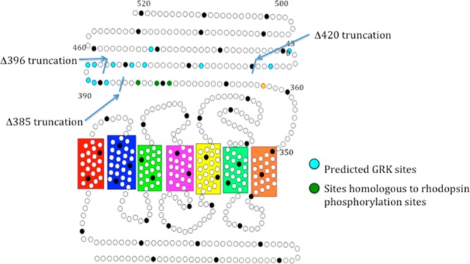
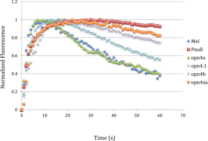
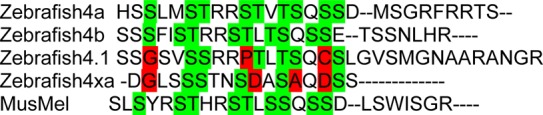
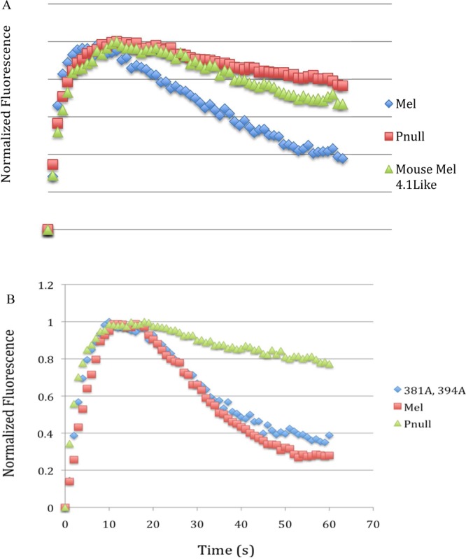
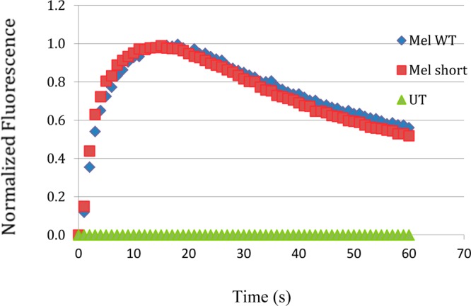
Light-activated opsins undergo carboxy-terminal phosphorylation, which contributes to the deactivation of their photoresponse. The photopigment melanopsin possesses an unusually long carboxy tail containing 37 serine and threonine sites that are potential sites for phosphorylation by a G-protein dependent kinase (GRK). Here, we show that a small cluster of six to seven sites is sufficient for deactivation of light-activated mouse melanopsin. Surprisingly, these sites are distinct from those that regulate deactivation of rhodopsin. In zebrafish, there are five different melanopsin genes that encode proteins with distinct carboxy-terminal domains. Naturally occurring changes in the same cluster of phosphorylatable amino acids provides diversity in the deactivation kinetics of the zebrafish proteins. These results suggest that variation in phosphorylation sites provides flexibility in the duration and kinetics of melanopsin-mediated light responses.


Figures

Figure 1
Figure 1
Secondary structure of mouse melanopsin. Each circle represents an amino acid, and every 10th amino acid is shaded black. Blue residues represent putative GRK phosphorylation sites (predicted by the group-based phosphorylation scoring (GPS) algorithm in GPS 2.0), green residues represent the homologous residues to those most often phosphorylated in rhodopsin, and the yellow dot represents a potential palmitoylation site.
Figure 2
Figure 2
Calcium imaging of carboxy-tail-truncation mutants. Kinetics of the calcium response of a series of carboxy-tail-truncation mutants compared to wild-type melanopsin and a phospho-null mutant. The fluorescent Ca2+-imaging data presented in this article is normalized to facilitate comparison of different melanopsin constructs. Supporting Information Figure 1 demonstrates that the normalized kinetics are not a function of the expression levels of the heterologously expressed melanopsin gene.
Figure 3
Figure 3
Sequence of deactivation control region. (A) Sites highlighted in green are predicted GRK family phosphorylation sites by the group-based phosphorylation algorithm. (B) Mutation of all phosphorylatable residues between amino acids 385 and 396 recapitulates the phospho-null phenotype.
Figure 4
Figure 4
Single phosphorylation site mutagenesis of putative deactivation control region. Each serine and threonine within the identified control region was mutated individually to an alanine. Deactivation of each mutant was determined in a kinetic calcium assay and compared to wild-type melanopsin. None of the six single mutations had any effect on signaling kinetics.
Figure 5
Figure 5
Kinetic calcium assay of zebrafish melanopsins expressed in HEK293 cells. Four of the five melanopsins found expressed in zebrafish were assayed for their deactivation kinetics. Opn4a and Opn4b were found to have similar deactivation kinetics to mouse melanopsin. Opn4.1 and Opn4xa were found to have extended deactivation kinetics matching the mouse melanopsin mutant lacking all carboxy-tail phosphorylaiton sites (phospho-null).
Figure 6
Figure 6
Alignment of zebrafish melanopsins with mouse melanopsin. Alignment of the zebrafish and mouse melanopsin sequences in the identified control region. Shown in green are the phosphorylation sites that are the same as mouse melanopsin, whereas the sites that are divergent from mouse melanopsin are in red.
Figure 7
Figure 7
Kinetic calcium assay of mouse melanopsin constructed to mimick zebrafish melanopsin opn4.1. (A) Kinetic calcium imaging of a mutant of mouse melanopsin engineered to match the pattern of phosphorylatable sites found in slow deactivating zebrafish melanopsin. (B) Mutation of the two conserved changed residues between opn4.1 and opn4xa and mouse melanopsin has no effect on signaling.
Figure 8
Figure 8
Calcium imaging of the two splice variants of mouse melanopsin. The long (Opn4L) (Mel WT) and short (Opn4S) (Mel short) isoforms of mouse melanopsins were assayed for their deactivation kinetics. They were found to have similar deactivation kinetics.

References

    1. Panda S.; Provencio I.; Tu D. C.; Pires S. S.; Rollag M. D.; Castrucci A. M.; Pletcher M. T.; Sato T. K.; Wiltshire T.; Andahazy M.; Kay S. A.; Van Gelder R. N.; Hogenesch J. B. (2003) Melanopsin is required for non-image-forming photic responses in blind mice. Science 301, 525–527. - PubMed
    1. Lucas R. J.; Hattar S.; Takao M.; Berson D. M.; Foster R. G.; Yau K. W. (2003) Diminished pupillary light reflex at high irradiances in melanopsin-knockout mice. Science 299, 245–247. - PubMed
    1. Tsai J. W.; Hannibal J.; Hagiwara G.; Colas D.; Ruppert E.; Ruby N. F.; Heller H. C.; Franken P.; Bourgin P. (2009) Melanopsin as a sleep modulator: Circadian gating of the direct effects of light on sleep and altered sleep homeostasis in Opn4–/– mice. PLoS Biol. 7, e1000125-1–e1000125-12. - PMC - PubMed
    1. LeGates T. A.; Altimus C. M.; Wang H.; Lee H. K.; Yang S.; Zhao H.; Kirkwood A.; Weber E. T.; Hattar S. (2012) Aberrant light directly impairs mood and learning through melanopsin-expressing neurons. Nature 491, 594–598. - PMC - PubMed
    1. Do M. T.; Kang S. H.; Xue T.; Zhong H.; Liao H. W.; Bergles D. E.; Yau K. W. (2009) Photon capture and signalling by melanopsin retinal ganglion cells. Nature 457, 281–287. - PMC - PubMed

Publication types