Laparoscopic surgery for double gastrointestinal stromal tumor of the stomach: a report of two cases
- PMID: 24678982
- PMCID: PMC3984392
- DOI: 10.1186/1477-7819-12-76
Laparoscopic surgery for double gastrointestinal stromal tumor of the stomach: a report of two cases
Abstract
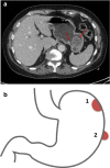
Gastrointestinal stromal tumors (GISTs) are mesenchymal tumors that originate from interstitial cells of Cajal or their stem cell-like precursors. Generally, GISTs have specific c-KIT gene mutations. The incidence of GISTs is estimated to be 10 to 20 cases/one million individuals, and GISTs typically affect people over 50 years of age. The majority of GISTs are solitary. However, multifocal GISTs have been observed, especially in children. We report on two unusual adult cases of double GISTs that were treated by laparoscopic surgery. The first patient presented a polypoid mass of the fundus and a second isolated smaller tumor in the posterior wall of the lesser curvature of the stomach. A histopathological examination confirmed that both tumors were GISTs and were c-KIT-positive. A total laparoscopic gastrectomy was performed. In the second patient, GISTs were identified at the level of the fundus and the greater curvature of the stomach. A laparoscopic partial sleeve gastrectomy was performed. Both surgeries were successful with no complications or relapses at three to five years following surgery.
Figures


References
-
- Miettinen M, Lasota J. Gastrointestinal stromal tumors: review on morphology, molecular pathology, prognosis, and differential diagnosis. Arch Pathol Lab Med. 2006;130:1466–1478. - PubMed
-
- Demetri GD, von Mehren M, Antonescu CR, DeMatteo RP, Ganjoo KN, Maki RG, Pisters PW, Raut CP, Riedel RF, Schuetze S, Sundar HM, Trent JC, Wayne JD. NCCN Task Force report: update on the management of patients with gastrointestinal stromal tumors. J Natl Compr Canc Netw. 2010;8(Suppl 2):S1–S41. quiz S42-44. - PMC - PubMed
Publication types
MeSH terms
LinkOut - more resources
Full Text Sources
Other Literature Sources
Medical

