. 2014 Sep;143(1):109-19.
doi: 10.1111/imm.12293.

CXCR3 deficiency enhances tumor progression by promoting macrophage M2 polarization in a murine breast cancer model

Affiliations

CXCR3 deficiency enhances tumor progression by promoting macrophage M2 polarization in a murine breast cancer model

Steve Oghumu et al. Immunology. 2014 Sep.

Abstract

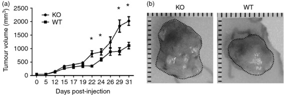
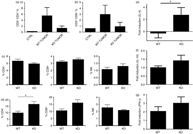
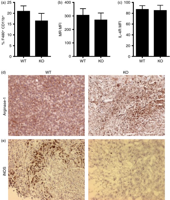
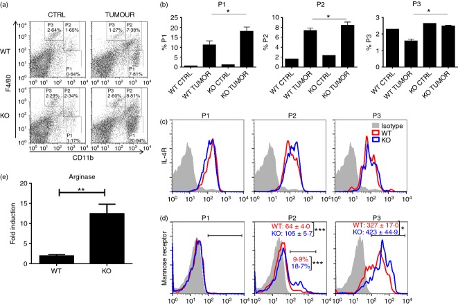
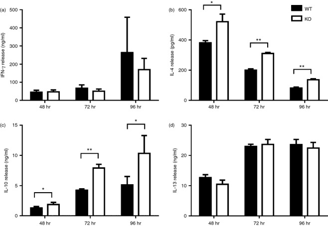
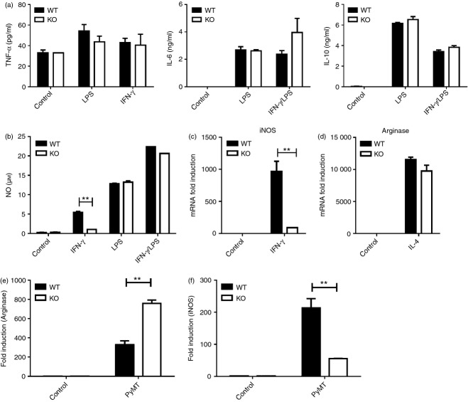
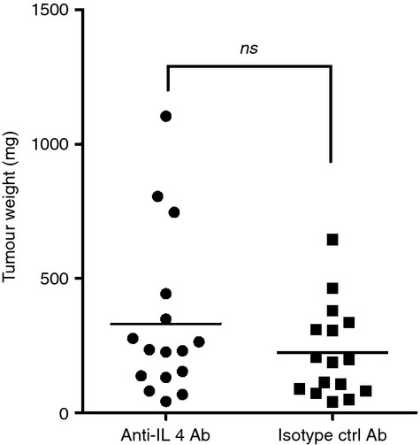
Tumor associated macrophages play a vital role in determining the outcome of breast cancer. We investigated the contribution of the chemokine receptor CXCR3 to antitumor immune responses using a cxcr3 deficient mouse orthotopically injected with a PyMT breast cancer cell line. We observed that cxcr3 deficient mice displayed increased IL-4 production and M2 polarization in the tumors and spleens compared to WT mice injected with PyMT cells. This was accompanied by larger tumor development in cxcr3(-/-) than in WT mice. Further, tumor-promoting myeloid derived immune cell populations accumulated in higher proportions in the spleens of cxcr3 deficient mice. Interestingly, cxcr3(-/-) macrophages displayed a deficiency in up-regulating inducible nitric oxide synthase after stimulation by either IFN-γ or PyMT supernatants. Stimulation of bone marrow derived macrophages by PyMT supernatants also resulted in greater induction of arginase-1 in cxcr3(-/-) than WT mice. Further, cxcr3(-/-) T cells activated with CD3/CD28 in vitro produced greater amounts of IL-4 and IL-10 than T cells from WT mice. Our data suggests that a greater predisposition of cxcr3 deficient macrophages towards M2 polarization contributes to an enhanced tumor promoting environment in cxcr3 deficient mice. Although CXCR3 is known to be expressed on some macrophages, this is the first report that demonstrates a role for CXCR3 in macrophage polarization and subsequent breast tumor outcomes. Targeting CXCR3 could be a potential therapeutic approach in the management of breast cancer tumors.

Keywords: CXCR3; arginase-1; breast cancer; inducible nitric oxide synthase; tumor associated macrophages.


Figures

Figure 1
Figure 1
Increased tumor growth in cxcr3−/− mice injected with PyMT breast cancer cell line: (a) Volumes of tumors of WT and cxcr3−/− mice injected with PyMT cancer cell line. Measurements were taken using an external digital caliper. Data are mean values from 8 to 10 individual mice per group and are representative of three independent experiments with similar results. *P < 0·05. (b) Representative images of tumors from WT and cxcr3−/− mice injected with PyMT cell line.
Figure 2
Figure 2
Frequency of tumor infiltrating lymphocytes in WT and cxcr3−/− C57BL/6 mice injected with PyMT breast cancer cell line: (a and b) Percentages of (a) CD4+ and (b) CD8+ T cells in tumors of WT and cxcr3−/− mice injected with PyMT cancer cells. (c and d) Percentages of CD4+ T cells, CD8+ T cells and NK cells in (c) spleens and (d) lymph nodes of WT and cxcr3−/− mice injected with PyMT cancer cells. Data are mean values from 4 to 5 individual mice per group and are representative of three independent experiments with similar results. *P < 0·05. (e–g) Real time PCR analysis of (e) IL-4, (f) IL-10 and (g) IFN-γ mRNA induction in tumors of WT and cxcr3−/− mice injected with PyMT cancer cells. Data shown are mean ± SE of duplicates from four or five individual mice per group and are representative of three individual experiments. *P < 0·05.
Figure 3
Figure 3
M2 macrophage populations are increased in cxcr3−/− mice containing tumors: (a) Frequency of F4/80+ CD11b+ macrophages in tumors of WT and cxcr3−/− mice injected with PyMT cancer cells. (b and c) Mean fluorescence intensities (MFIs) of (b) mannose receptor and (c) IL-4 receptor on F4/80+ CD11b+ macrophages in breast tumors of WT and cxcr3−/− mice injected with PyMT cells. All flow cytometric data shown are mean ± SE of four or five individual mice per group and are representative of three independent experiments. (d and e) Representative microscopic images showing immunohistochemical staining of (d) arginase-1 and (e) iNOS in breast cancer tumor sections from WT and cxcr3−/− mice injected with PyMT cells.
Figure 4
Figure 4
Accumulation of tumor promoting myeloid derived immune cell populations in the spleens of cxcr3−/− mice with breast cancer: (a) Representative dot plots showing myeloid derived immune cell populations in the spleens of PyMT injected WT and cxcr3−/− mice. Splenocytes were stained with CD11b, F4/80, CD206 and IL-4R. (b) Bar graph showing frequency of various myeloid derived immune cell populations in spleens of WT and cxcr3−/− mice injected with PyMT cancer cells, based on the gating scheme depicted in Fig. a. Data shown are mean ± SE of four or five individual mice per group and are representative of three individual experiments with similar results. *P < 0·05 (c) Representative histogram plots showing expression of IL-4 receptor on P1, P2 and P3 gated populations in spleens of WT and cxcr3−/− mice injected with PyMT cancer cells. (d) Representative histogram plots showing expression of Mannose receptor (CD206) on P1, P2 and P3 gated populations in spleens of WT and cxcr3−/− mice injected with PyMT cancer cells. Numbers represent average of mean fluorescence intensities (MFIs) of mannose receptor expression or percentages of mannose receptor expressing cells on gated populations. Data shown are mean ± SE of four or five individual mice per group and are representative of three independent experiments. (e) Real time PCR analysis of arginase-1 mRNA induction in spleens of PyMT injected WT and cxcr3−/− mice **P < 0·01.
Figure 5
Figure 5
In vitro activation of T cells favor Th2 polarization in cxcr3−/− mice: (A - D) Cytokine production of T cells from naïve WT and cxcr3−/− mice activated with plate bound anti CD3/CD28 antibodies. (a) IFN-γ, (b) IL-4, (c) IL-10 and (d) IL-13 production were measured in culture supernatants by ELISA. Data shown are mean ± SE of duplicates from three or four individual mice per group and are representative of two independent experiments *P < 0·05, **P < 0·01.
Figure 6
Figure 6
In vitro activation of macrophages favor M2 polarization in cxcr3−/− mice: (a) Cytokine production by BMDMs from WT and cxcr3−/− mice stimulated in vitro by LPS, IFN-γ and/or LPS/IFN-γ as determined by ELISA. Data shown are mean ± SE of duplicates from three or four individual mice per group and are representative of two independent experiments. (b) Nitric oxide production by BMDMs from WT and cxcr3−/− mice stimulated in vitro by IFN-γ for 48 hr, as determined by Griess reaction. (c) Real time PCR analysis of inducible nitric oxide synthase (iNOS) mRNA induction in BMDMs from naïve WT and cxcr3−/− mice stimulated with IFN-γ. (d) Real time PCR analysis of arginase-1 mRNA induction in BMDMs from naïve WT and cxcr3−/− mice stimulated with IL-4. (E and F) Real time PCR analysis of (e) arginase-1 and (f) iNOS mRNA induction in BMDMs from naïve WT and cxcr3−/− mice stimulated with PyMT cancer cell culture supernatants. Data shown are mean ± SE of duplicates from three or four individual mice per group and are representative of three independent experiments **P < 0·01.
Figure 7
Figure 7
IL-4 blockade does not reduce tumor progression in cxcr3−/− mice: Tumor weights of cxcr3−/− mice treated with anti-IL-4 antibody or isotype control antibody. Tumors weights were measured 5 weeks after injection of PyMT cells. Data shown are mean values from fifteen to eighteen individual mice per group from 3 independent experiments (ns – not significant).

References

    1. Sato E, Olson SH, Ahn J, et al. Intraepithelial CD8+ tumor-infiltrating lymphocytes and a high CD8+/regulatory T cell ratio are associated with favorable prognosis in ovarian cancer. Proc Natl Acad Sci U S A. 2005;102:18538–43. - PMC - PubMed
    1. Galdiero MR, Garlanda C, Jaillon S, Marone G, Mantovani A. Tumor associated macrophages and neutrophils in tumor progression. J Cell Physiol. 2013;228:1404–12. - PubMed
    1. Menard S, Tomasic G, Casalini P, et al. Lymphoid infiltration as a prognostic variable for early-onset breast carcinomas. Clin Cancer Res. 1997;3:817–9. - PubMed
    1. DeNardo DG, Coussens LM. Inflammation and breast cancer. Balancing immune response: crosstalk between adaptive and innate immune cells during breast cancer progression. Breast Cancer Res. 2007;9:212. - PMC - PubMed
    1. Sica A, Mantovani A. Macrophage plasticity and polarization: in vivo veritas. J Clin Investig. 2012;122:787–95. - PMC - PubMed

Publication types

MeSH terms