Review
. 2014 Apr:27:32-43.
doi: 10.1016/j.ceb.2013.11.001. Epub 2013 Nov 28.

Structural approaches to understanding retinal proteins needed for vision

Affiliations
Review

Structural approaches to understanding retinal proteins needed for vision

Tivadar Orban et al. Curr Opin Cell Biol. 2014 Apr.

Abstract

The past decade has witnessed an impressive expansion of our knowledge of retinal photoreceptor signal transduction and the regulation of the visual cycle required for normal eyesight. Progress in human genetics and next generation sequencing technologies have revealed the complexity behind many inherited retinal diseases. Structural studies have markedly increased our understanding of the visual process. Moreover, technical innovations and improved methodologies in proteomics, macromolecular crystallization and high resolution imaging at different levels set the scene for even greater advances. Pharmacology combined with structural biology of membrane proteins holds great promise for developing innovative accessible therapies for millions robbed of their sight or progressing toward blindness.


Figures

Fig. 1
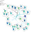
Fig. 1. Phylogenetic tree of GPCRs with known X-ray structures
Branches and sub-branches (detailed methods are described elsewhere) of the GPCR phylogenetic tree are shown (adapted from Katritch et al. [25]). Branches of varying thickness represent different subgroups. Known structures from class A (rhodopsin subfamily, light blue branches), class B (secretin subfamily, red branches), and class F (frizzled, green branches) GPCRs are shown, but other groups such as the adhesion and glutamate GPCRs are not included. Structures of known GPCRs obtained in the presence of different ligands are clustered together. Types of ligands are identified by geometrical shapes placed at the top left corner of each individual GPCR structure. Structures with partial agonists are denoted with open triangles, agonists are highlighted with closed triangles, antagonists are identified by open squares, partial inverse agonists are marked by closed squares and inverse agonists are shown with closed circles. GPCRs bound to different inhibitors are identified by their gene names. B-factors for each individual GPCR structure were used to generate the GPCR rainbow color coding (blue through red, for minimum and maximum B-factor values, respectively). The gene names of the clustered structures and their corresponding PDB codes are: RHO (1U19 and 3CAP); ADRB1 (2VT4, 2Y02, and 2Y01); ADRB2 (2R4R and 2RH1); HTR2B (4IB4); HTR1B (4IAQ); CHRM3 (4DAJ); CHRM2 (3UON); DRD3 (3PBL); HRH1 (3RZE); F2R (3VW7); CXCR4 (3ODU); OPRD1 (4EJ4); OPRM1 (4DKL); OPRK1 (4DJH); OPRL1 (4EA3); NTSR1 (4GRV); ADORA2A (3QAK; 3VGA), and 3EML); EDG1 (3V2W); CRF1R (4K5Y); GCGR (4L6R); SMOH (4JKV).
Fig. 2
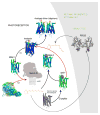
Fig. 2. Structures of rhodopsin, opsin and activated intermediates determined by X-ray crystallography
Color coding of the shown structures was derived from B- factors of the αC atoms as noted in Fig. 1. Ground state rhodopsin (with bound 11-cis-retinal depicted as a red sphere) is activated by a photon of light. Prepared under different experimental conditions, presented GPCR structures were superimposed on ground state rhodopsin and generated using PDB codes: 1U19, 3C9l, 1GZM, 2I35, and 2I36. Photoisomerization of 11-cis-retinal to all-trans-retinal (shown as orange spheres) is accompanied by conformational changes in the transmembrane helices together with a variety of local environmental changes (such as rearrangement of the internal water molecular network). Some of these changes are reflected in B-factor values. The Meta II structure (PDB code 3PXO) contains the all-trans-retinal chromophore. A potential structure of the Meta II-Gt complex is shown in dark grey and light grey shades and indicated by red arrows. The structures of both opsin* and opsin* with bound Gt peptide lack a visual chromophore (PDB codes 3CAP (in gray cartoons because the B-factors for this structure are incorrect) and 3DQB, respectively) and both can be converted to a Meta II state [14]) in the presence of all-trans-retinal (orange sticks) (black arrows, PDB code 3PQR). Opsin structures are not apo-receptors, because they contained bound detergent molecules (depicted in red) [90]. The Gt peptide is represented as grey surface. However, addition of 11-cis-retinal (red sticks) to opsin* crystals cannot successfully regenerate ground state rhodopsin (red arrow). RPE65 (4F3D), a component of the visual cycle, is shown as a dark gray cartoon on a light gray background denoting the retinal pigmented epithelium (RPE). Recycling of all-trans-retinal to 11-cis-retinal through the visual cycle is indicated by gray arrows. Rhodopsin has propensity to oligomerize in both native and expression systems [–93].
Fig. 3
Fig. 3. The β2-adrenergic-Gs complex modeled into a phospholipid membrane bilayer
The β2-adrenergic receptor in complex with Gs (PDB code: 3SN6) is inserted in a lipid bilayer. The phospholipid units (1-palmitoyl-2-oleoyl-sn-glycero-3-phosphocholine) of the lipid bilayer are depicted as gray spheres and the model has been described in detail elsewhere [51]. The position of the β2-adrenergic transmembrane domain (light blue cartoon) along the z axis of the phospholipid bilayer was chosen by superposition with rhodopsin (not shown). The position of rhodopsin (PDB code: 1U19) in turn is presented such that the palmitoyl fatty acyl chains attached to residues Cys322 and Cys323 are positioned at the same depth as the phospholipid fatty acyl chains of the membrane leaflet. Both the Nb35 nanobody and T4 lysozyme [49] are manifested as light gray structures. The Gs domains are represented as follows: G and G are shown as cyan cartoons whereas G is pictured as a blue cartoon. Amino acid residues (Asn121 to Leu153) of the G domain that overlap with the phospholipid head group region of the bilayer model are shown as a red cartoon. The face-on view of the transmembrane region is presented as a z-plane section of the phospholipid bilayer.
Fig. 4
Fig. 4. Orientation of GPCR-G protein complexes in a phospholipid membrane
Semi-empirical models of the complex formed between light-activated rhodopsin dimer and a Gt heterotrimer (A and B) [52] and the T4L-β2AR-Gs-nanobody complex (C) [48] were fitted into a 3D molecular envelope calculated from projections of negatively stained, bis[succinimidyl] 2,2,4,4-glutarate (DSG) crosslinked rhodopsin*-Gt complexes purified in lauryl maltose neopentyl glycol (LMNG). Fitting of the rhodopsin*-Gt model generated with the structure of inactive Gt (PDB code: 1GOT) leaves a significant unoccupied density above one of the rhodopsin molecules (A), which becomes occupied after a 30o hinge-like motion of the α-helical domain is applied (B). Though fitting the T4L-β2-adrenergic receptor-Gs-nanobody structure into our EM 3D map leaves sufficient space to accommodate a second molecule of this receptor, conformation of the G helical domain is inconsistent with our EM-density (C). Thus, the favored structure of the rhodopsin-Gt complex appears to be that shown in (B). Photoactivated rhodopsin (Rho*) that binds the C-terminal peptide derived from G and the T4L-β2-adrenergic receptor-nanobody molecule are both depicted in yellow. The second rhodopsin molecule in (A) and (B) is shown in gray. G, G, G are colored pink, dark cyan and dark blue, respectively.

References

    1. Mustafi D, Maeda T, Kohno H, Nadeau JH, Palczewski K. Inflammatory priming predisposes mice to age-related retinal degeneration. J Clin Invest. 2012;122:2989–3001. - PMC - PubMed
    1. Arshavsky VY, Burns ME. Photoreceptor signaling: supporting vision across a wide range of light intensities. J Biol Chem. 2012;287:1620–1626. - PMC - PubMed
    1. Palczewski K. Chemistry and biology of vision. J Biol Chem. 2012;287:1612–1619. Visual perception in humans is initiated through absorption of photons by photoreceptors in the retina, a layered sensory organ containing all the necessary functional and structural proteins to support this process. How this remarkable tissue develops and operates over such an incredible dynamic range and how retinoids are recycled are some of the most fascinating questions in biology. With current methodology, it is now possible to identify all components of the retina and trace mutations to multiple blinding diseases. - PMC - PubMed
    1. Gilliam JC, Chang JT, Sandoval IM, Zhang Y, Li T, Pittler SJ, Chiu W, Wensel TG. Three-dimensional architecture of the rod sensory cilium and its disruption in retinal neurodegeneration. Cell. 2012;151:1029–1041. Using cryo-electron tomography, Wensel and colleagues obtained for the first time the 3D structure of the connecting cilium and adjacent cellular structures of a modified primary cilium in the rod outer segment isolated from wild type and disease model mice. This work revealed new structural features involved in cellular transport that is impaired by the involved genetic defects. - PMC - PubMed
    1. Palczewski K, Kumasaka T, Hori T, Behnke CA, Motoshima H, Fox BA, Le Trong I, Teller DC, Okada T, Stenkamp RE, Yamamoto M, Miyano M. Crystal structure of rhodopsin: A G protein-coupled receptor. Science. 2000;289:739–745. - PubMed

Publication types

MeSH terms