Review
. 2014 Aug:27:1-7.
doi: 10.1016/j.sbi.2014.02.005. Epub 2014 Mar 25.

Single particle electron cryo-microscopy of a mammalian ion channel

Affiliations
Review

Single particle electron cryo-microscopy of a mammalian ion channel

Maofu Liao et al. Curr Opin Struct Biol. 2014 Aug.

Abstract

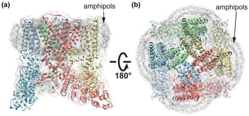
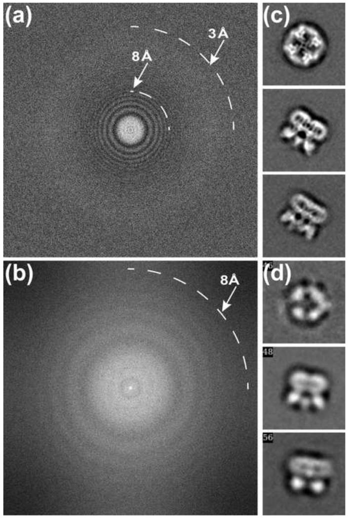
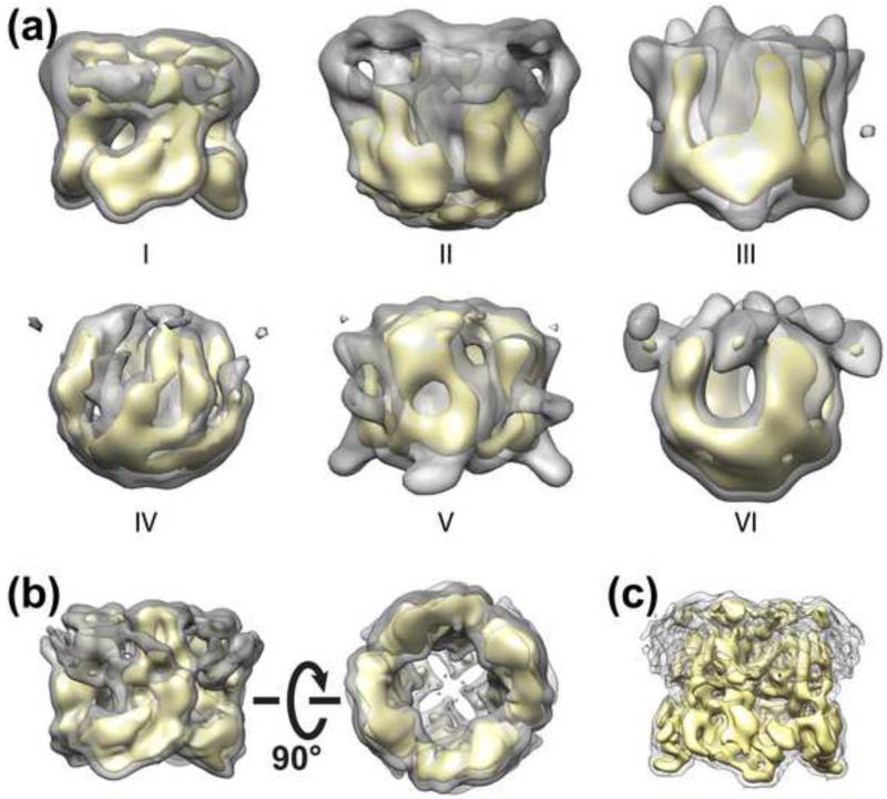
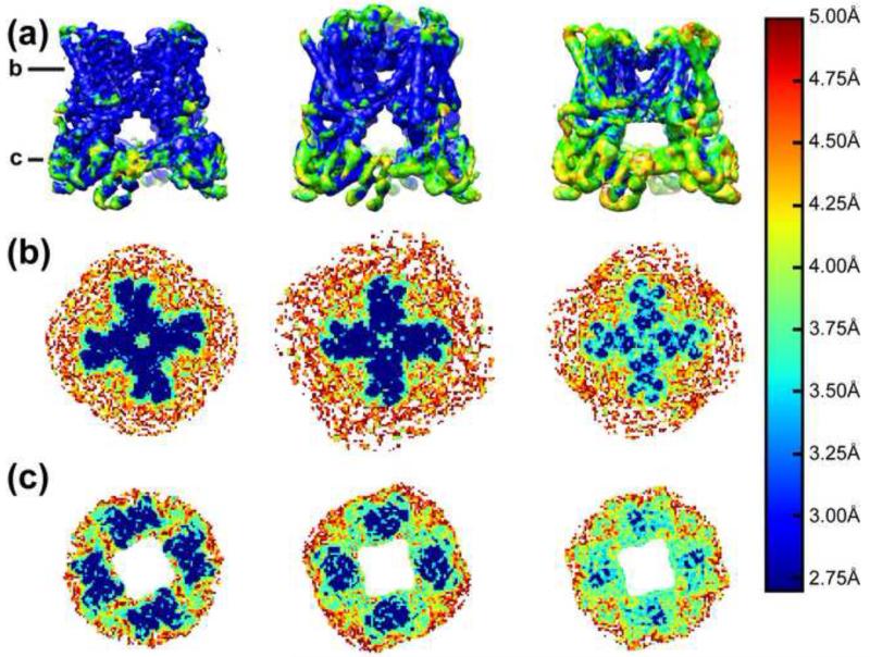
The transient receptor potential (TRP) ion channel family is large and functionally diverse, second only to potassium channels. Despite their prominence within the animal kingdom, TRP channels have resisted crystallization and structural determination for many years. This barrier was recently broken when the three-dimensional structure of the vanilloid receptor 1 (TRPV1) was determined by single particle electron cryo-microscopy (cryo-EM). Moreover, this is the first example in which the near atomic resolution structure of an integral membrane protein was elucidated by this technique and in a manner not requiring crystals, demonstrating the transformative power of single particle cryo-EM for revealing high-resolution structures of integral membrane proteins, particularly those of mammalian origin. Here we summarize technical advances, in both biochemistry and cryo-EM, that led to this major breakthrough.


Figures

Figure 1
Figure 1. Structure of rat TRPV1 in amphipols
(a) Side and (b) top views of the structure of rat TRPV1 in ribbon diagram. The density of amphipols is marked.
Figure 2
Figure 2. Cryo-EM data are significantly improved with direct detection camera and motion correction
(a) Fourier transform of a typical cryo-EM image of TRPV1 ion channel embedded in thin layer of vitreous ice recorded with the direct detection camera, K2 Summit, after motion correction. Thon rings are visible up to ~3Å resolution. The position corresponding to 8Å is also marked. (b) Fourier transform of a similar image recorded using a phosphor scintillator based CMOS camera, TemF816. Thon rings are visible up to ~8Å resolution. (c) Three typical 2D class averages of images recorded with the K2 camera. (d) Similar 2D class averages of TRPV1 particle images recorded with the TemF816.
Figure 3
Figure 3. 3D classification of particle images of the TRPV1 ion channel
(a) A total of 45,625 particle images were classified into six classes. 3D reconstructions calculated for each class are shown. The number of particles in each class is: 10,357 (I); 9,527 (II); 3,778 (III); 11,671 (IV); 5,328 (V); 4,964 (VI). Only the first class can be refined to high resolution, 8.8Å. (b) Two different views of the 3D reconstruction calculated from the entire dataset. The nominal resolution is ~9.3Å, but the center of the transmembrane domain is hollow, showing no sign of transmembrane helices. (c) The final 3D reconstruction calculated from class I at a nominal resolution of 8.8Å, showing all transmembrane helices.
Figure 4
Figure 4. Estimation of local resolution
(a) Local resolution of 3D reconstructions of TRPV1 (left), in complex with RTX/DkTx (middle) and capsaicin (right). Local resolutions are color-coded. The color scale is shown in the right side. (b) and (c) are two slides (marked in (a)) perpendicular to the 4-fold symmetry axis.

References

    1. Carpenter EP, Beis K, Cameron AD, Iwata S. Overcoming the challenges of membrane protein crystallography. Curr Opin Struct Biol. 2008;18:581–586. - PMC - PubMed
    1. Fujiyoshi Y. Future directions of electron crystallography. Methods Mol Biol. 2013;955:551–568. - PubMed
    1. Loll PJ. Membrane protein structural biology: the high throughput challenge. J Struct Biol. 2003;142:144–153. - PubMed
    1. Grigorieff N, Harrison SC. Near-atomic resolution reconstructions of icosahedral viruses from electron cryo-microscopy. Curr Opin Struct Biol. 2011;21:265–273. - PMC - PubMed
    1. Lau WC, Rubinstein JL. Subnanometre-resolution structure of the intact Thermus thermophilus H+-driven ATP synthase. Nature. 2012;481:214–218. 3D reconstruction of H+-driven ATP synthase was the first membrane protein structure determined by single particle cryo-EM that reached a resolution of better than 10Å. Transmembrane helices are resolved int this 3D reconstruction.

Publication types

Substances