. 2014 Jul;61(1):107-15.
doi: 10.1016/j.jhep.2014.03.028. Epub 2014 Mar 27.

Differential regulation of inflammation and apoptosis in Fas-resistant hepatocyte-specific Bid-deficient mice

Affiliations

Differential regulation of inflammation and apoptosis in Fas-resistant hepatocyte-specific Bid-deficient mice

Milos Lazic et al. J Hepatol. 2014 Jul.

Abstract

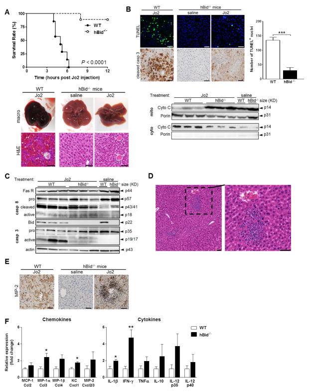
Background & aims: Activation of Fas death receptor results in apoptosis in multiple organs, particularly liver, in a process dependent on Bid cleavage. Mice injected with an anti-Fas antibody die within hours of acute liver failure associated with massive apoptosis and hemorrhage. Our aim was to investigate the crosstalk of apoptotic and inflammatory pathways and the contribution of selective hepatocellular apoptosis during in vivo Fas activation.

Methods: We generated hepatocyte-specific Bid deficient mice (hBid(-/-)). Acute liver injury was induced by Fas-activating antibody (Jo2) in a time-course study.

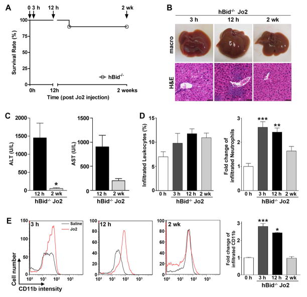
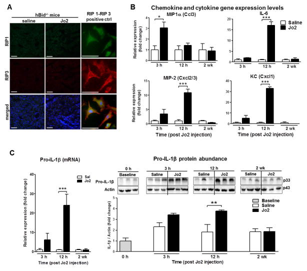
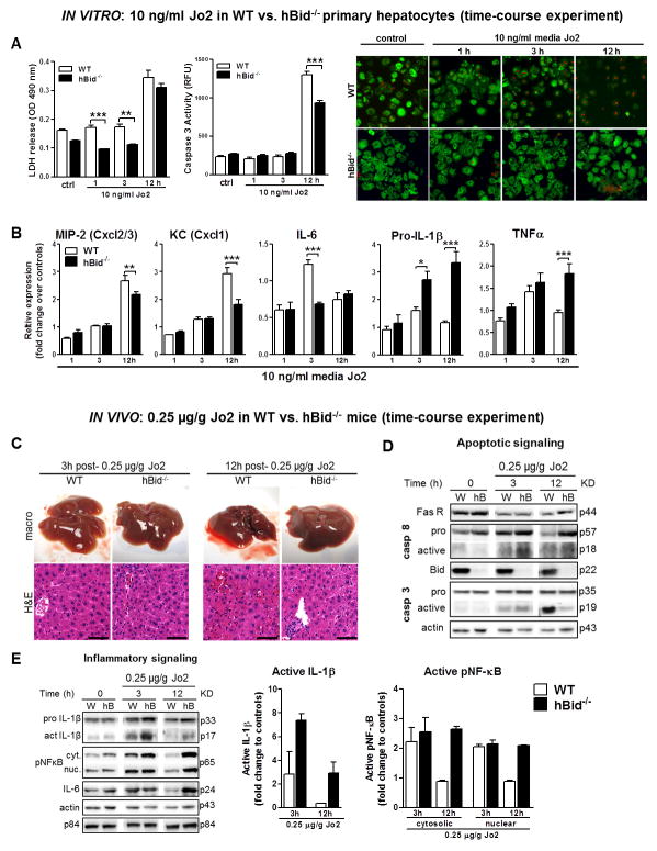
Results: In contrast to controls, nearly all Jo2 injected hBid(-/-) survived. Their livers showed complete protection against hepatocellular apoptosis with minimal focal hemorrhagic changes and mainly non-parenchymal cell apoptosis. In agreement, the hepatocytes had no mitochondrial cytochrome c release in cytosol, or caspase 3 activation. hBid(-/-) livers showed marked increase in acute inflammatory foci composed of neutrophils and monocytes associated with the increased expression of proinflammatory chemokines and cytokines, in the manner dependent on non-canonical interleukin-1β activation and amplified in the absence of caspase-3 activation. In addition, hBid(-/-) mice were completely protected from hepatotoxicity and the infiltrated cells were cleared 2 weeks post single Jo2 injection.

Conclusions: Hepatocyte Bid suppression is critical for the resistance to the lethal effects of Fas activation in vivo. Fas signaling induces differential activation of non-canonical interleukin-1β maturation, amplified in the absence of apoptotic Bid-mitochondrial loop, in hepatocytes. These findings may have important pathophysiological and therapeutic implications in a variety of liver disorders associated with Fas activation.

Keywords: Acute liver failure; Crosstalk of inflammatory and apoptotic pathways; Fas-mediated hepatic apoptosis; Hepatoprotection; Inflammation; Non-canonical IL-1β maturation.


Conflict of interest statement

Conflict of Interest: Authors have nothing to disclose.

Figures

Figure 1
Figure 1. hBid−/− mice are resistant to Fas-induced mortality, hepatocyte apoptosis and gross liver hemorrhage, but are prone to the formation of marked acute inflammatory foci and neutrophilic infiltration
(A) Survival curve of wild-type (WT; filled circles, n = 7) and hBid−/− mice (circles, n = 9) in response to a single IP injection of 100 μg Jo2 over the period of 12 h post injection, P < 0.0001. Mice were sacrificed 12 h after Jo2-injection or shortly before death. Liver macro pictures and haematoxylin-eosin staining of liver sections of Jo2 injected WT mice and vehicle control (saline) or Jo2 injected hBid−/− mice, respectively. Note the marked disruption of liver architecture, massive hepatic apoptosis and gross hemorrhage due to Fas activation in WT mice. (B) DNA fragmentation by TUNEL assay and detection of apoptosis by immunohistochemical staining specific for cleaved caspase 3 in WT and vehicle control or Jo2 injected hBid−/− mice. Quantification of the representative 40x TUNEL fields of WT (n = 7) vs. hBid−/− (n = 5) mice. Assessment of cytochrome c distribution in mitochondria vs. cytosol of fractionated liver lysates in WT and hBid−/− mice. (C) Analysis of the activation of initiator caspase 8 and effector caspase 3 in livers by immunoblots after injection of Jo2 in WT and vehicle control or Jo2 injected hBid−/− mice. Representative blots show the activated fragments: p18 of caspase-8 and p17/19 of caspase-3, in addition to their pro-caspase counterparts. (D) Haematoxylin-eosin staining of liver sections of Jo2 injected hBid−/− mice, emphasizing neutrophilic infiltration; (E) immunohistochemical staining specific for MIP-2 in WT and vehicle control or Jo2 injected hBid−/− mice. Note the marked neutrophilic infiltration and the concomitant focal increase in the abundance of MIP-2 in hBid−/− compared to WT mice. (F) Expression profiling of chemokines and cytokines involved in Fas-mediated liver inflammation. All animals (n = 4, each group; WT, open bars; hBid−/−, black bars) were injected with Jo2. All gene expression levels are shown relative to the expression levels of WT mice. Values are mean ± SEM. *P < 0.05, **P < 0.01, ***P < 0.001 compared to controls. Scale bar, 50 μm.
Figure 2
Figure 2. Time-course study: resistance of hBid−/− mice to Fas-induced mortality and the complete resolution of inflammation two weeks post anti-Fas antibody injection
(A) Survival curve of hBid−/− mice (n = 10) in response to a single IP injection of 100 μg anti-Fas antibody (Jo2) over the period of 2 weeks. (B) Liver macro pictures and haematoxylin-eosin staining of liver sections of hBid−/− mice post Jo2-injection. (C) Plasma levels of ALT and AST of hBid−/− mice 12 h and 2 wk post Jo2-injection (hBid−/− n = 4). (D) Percent of leukocytes and fold change of neutrophils (respective to saline injections at each time point) infiltrated into the liver of hBid−/− post Jo2-injection by flow cytometry analysis. (E) Representative histograms of infiltrated monocytes (CD11b+). Red line: hBid−/− Jo2 injection, Black line: hBid−/− saline injection. Bar graph shows quantification of infiltrated cells as shown by fold change compared to saline injections at each time point. Values are mean ± SEM. *P < 0.05, **P < 0.01, ***P < 0.001 compared to baseline controls. Scale bar, 50 μm.
Figure 3
Figure 3. Time-course study: Fas mediates hepatocyte chemokine and IL-1β production in an RIP-independent manner
(A) Hepatic Fas signaling does not induce RIP-1/RIP-3 complex required for programmed necrosis 12 h post Jo2-injection. Immunofluorescent staining specific for RIP-1 and RIP-3 kinases and merged image in vehicle and control Jo2 injected hBid−/− mice. Necrotic MEFs were used as a positive control for the formation of RIP-1/RIP-3 complex. Scale bar, 50 μm. (B) Hepatocyte (at 3 h and 12 h) and whole liver (at 2 wk) chemokine gene expression levels in Fas-mediated liver inflammation of hBid−/− mice. Gene expression was measured in hBid−/− mice (n = 3, each group; saline, open bars; Jo2 treated, black bars) 3 h, 12 h and 2 wk post Jo2 injection. All gene expression levels were normalized to housekeeping control and shown relative to the expression levels of saline treated mice. (C) Hepatocyte pro-IL-1β gene expression and protein levels post Jo2-injections in Fas-mediated liver inflammation of hBid−/− mice (saline, open bars; Jo2 treated, black bars). Values are mean ± SEM. *P < 0.05, **P < 0.01, ***P < 0.001 compared to controls.
Figure 4
Figure 4. In vitro and in vivo low Jo2 dose time-course study dissociates apoptotic and inflammatory pathways
(A) The assessment of apoptosis and cell death in WT (open bars) and hBid−/− (black bars) primary hepatocytes. From left to right, lactate dehydrogenase (LDH) cytotoxicity assay, caspase 3 activity and live/dead cell viability assay (red stains nuclei of dead cells, green stains living cells). (B) Hepatocyte inflammatory chemokine and cytokine gene expression normalized to untreated controls (n=6 each group). (C) Liver macro pictures and haematoxylin-eosin staining of liver sections of Jo2 injected WT (open bars) and hBid−/− mice (black bars). Note the small levels of liver hemorrhage in WT and the formation of inflammatory foci hBid−/− mice at 12 h post Jo2 injection. (D) Representative immunoblots of select apoptotic signaling pathways in Jo2 injected WT and hBid−/− mice. (E) Representative immunoblots and protein levels of select inflammatory signaling pathways probed in cytosolic (cyt.) and nuclear (nuc.) fractions. Note the marked abundance of mature/active IL-1β at 3 h and sustained activation of NF-κB and the corresponding abundance of IL-6 through 12 h post Jo2-treatment in hBid−/− mice. W, WT; hB, hBid−/−. Values are mean ± SEM. *P < 0.05, **P < 0.01, ***P < 0.001 compared to controls. Scale bar, 50 μm.

Similar articles

Cited by

  • Bacteroides acidifaciens in the gut plays a protective role against CD95-mediated liver injury.
    Wang H, Wang Q, Yang C, Guo M, Cui X, Jing Z, Liu Y, Qiao W, Qi H, Zhang H, Zhang X, Zhao N, Zhang M, Chen M, Zhang S, Xu H, Zhao L, Qiao M, Wu Z. Wang H, et al. Gut Microbes. 2022 Jan-Dec;14(1):2027853. doi: 10.1080/19490976.2022.2027853. Gut Microbes. 2022. PMID: 35129072 Free PMC article.
  • Apoptotic cell death in disease-Current understanding of the NCCD 2023.
    Vitale I, Pietrocola F, Guilbaud E, Aaronson SA, Abrams JM, Adam D, Agostini M, Agostinis P, Alnemri ES, Altucci L, Amelio I, Andrews DW, Aqeilan RI, Arama E, Baehrecke EH, Balachandran S, Bano D, Barlev NA, Bartek J, Bazan NG, Becker C, Bernassola F, Bertrand MJM, Bianchi ME, Blagosklonny MV, Blander JM, Blandino G, Blomgren K, Borner C, Bortner CD, Bove P, Boya P, Brenner C, Broz P, Brunner T, Damgaard RB, Calin GA, Campanella M, Candi E, Carbone M, Carmona-Gutierrez D, Cecconi F, Chan FK, Chen GQ, Chen Q, Chen YH, Cheng EH, Chipuk JE, Cidlowski JA, Ciechanover A, Ciliberto G, Conrad M, Cubillos-Ruiz JR, Czabotar PE, D'Angiolella V, Daugaard M, Dawson TM, Dawson VL, De Maria R, De Strooper B, Debatin KM, Deberardinis RJ, Degterev A, Del Sal G, Deshmukh M, Di Virgilio F, Diederich M, Dixon SJ, Dynlacht BD, El-Deiry WS, Elrod JW, Engeland K, Fimia GM, Galassi C, Ganini C, Garcia-Saez AJ, Garg AD, Garrido C, Gavathiotis E, Gerlic M, Ghosh S, Green DR, Greene LA, Gronemeyer H, Häcker G, Hajnóczky G, Hardwick JM, Haupt Y, He S, Heery DM, Hengartner MO, Hetz C, Hildeman DA, Ichijo H, Inoue S, Jäättelä M, Janic A, Joseph B, Jost PJ, Kanneganti TD, Karin M, Kashkar H, Kaufmann T, Kelly … See abstract for full author list ➔ Vitale I, et al. Cell Death Differ. 2023 May;30(5):1097-1154. doi: 10.1038/s41418-023-01153-w. Epub 2023 Apr 26. Cell Death Differ. 2023. PMID: 37100955 Free PMC article. Review.
  • Liver Bid suppression for treatment of fibrosis associated with non-alcoholic steatohepatitis.
    Eguchi A, De Mollerat Du Jeu X, Johnson CD, Nektaria A, Feldstein AE. Eguchi A, et al. J Hepatol. 2016 Mar;64(3):699-707. doi: 10.1016/j.jhep.2015.11.002. Epub 2015 Nov 7. J Hepatol. 2016. PMID: 26555271 Free PMC article.
  • Transmembrane BAX Inhibitor motif-containing 1, a novel anti-inflammatory approach for nonalcoholic steatohepatitis treatment.
    Wree A, Inzaugarat ME, Feldstein AE. Wree A, et al. Hepatology. 2018 Jan;67(1):438-441. doi: 10.1002/hep.29495. Hepatology. 2018. PMID: 28859227 Free PMC article. No abstract available.
  • Bid-deficient fish delay grass carp reovirus (GCRV) replication and attenuate GCRV-triggered apoptosis.
    He L, Wang H, Luo L, Li Y, Huang R, Liao L, Zhu Z, Wang Y. He L, et al. Oncotarget. 2017 Jul 22;8(44):76408-76422. doi: 10.18632/oncotarget.19460. eCollection 2017 Sep 29. Oncotarget. 2017. PMID: 29100321 Free PMC article.

References

    1. Galle PR, Hofmann WJ, Walczak H, Schaller H, Otto G, Stremmel W, et al. Involvement of the CD95 (APO-1/Fas) receptor and ligand in liver damage. J Exp Med. 1995;182:1223–30. - PMC - PubMed
    1. Suda T, Takahashi T, Golstein P, Nagata S. Molecular cloning and expression of the Fas ligand, a novel member of the tumor necrosis factor family. Cell. 1993;75:1169–78. - PubMed
    1. Ogasawara J, Watanabe-Fukunaga R, Adachi M, Matsuzawa A, Kasugai T, Kitamura Y, et al. Lethal effect of the anti-Fas antibody in mice. Nature. 1993;364:806–9. - PubMed
    1. Itoh N, Yonehara S, Ishii A, Yonehara M, Mizushima S, Sameshima M, et al. The polypeptide encoded by the cDNA for human cell surface antigen Fas can mediate apoptosis. Cell. 1991;66:233–43. - PubMed
    1. Nagata S. Apoptosis by death factor. Cell. 1997;88:355–65. - PubMed

Publication types

MeSH terms