. 2014 May;10(5):400-6.
doi: 10.1038/nchembio.1496. Epub 2014 Mar 30.

Amphotericin forms an extramembranous and fungicidal sterol sponge

Affiliations

Amphotericin forms an extramembranous and fungicidal sterol sponge

Thomas M Anderson et al. Nat Chem Biol. 2014 May.

Abstract

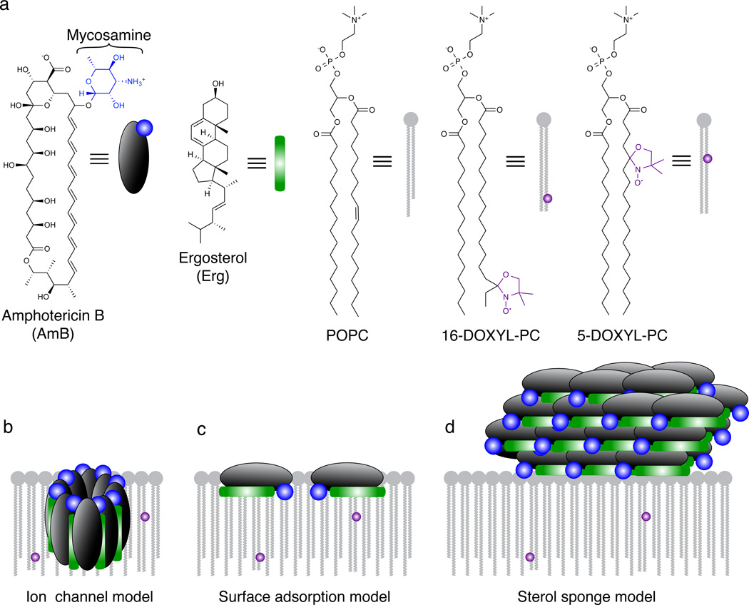
For over 50 years, amphotericin has remained the powerful but highly toxic last line of defense in treating life-threatening fungal infections in humans with minimal development of microbial resistance. Understanding how this small molecule kills yeast is thus critical for guiding development of derivatives with an improved therapeutic index and other resistance-refractory antimicrobial agents. In the widely accepted ion channel model for its mechanism of cytocidal action, amphotericin forms aggregates inside lipid bilayers that permeabilize and kill cells. In contrast, we report that amphotericin exists primarily in the form of large, extramembranous aggregates that kill yeast by extracting ergosterol from lipid bilayers. These findings reveal that extraction of a polyfunctional lipid underlies the resistance-refractory antimicrobial action of amphotericin and suggests a roadmap for separating its cytocidal and membrane-permeabilizing activities. This new mechanistic understanding is also guiding development of what are to our knowledge the first derivatives of amphotericin that kill yeast but not human cells.


Figures

Figure 1
Figure 1. Models for the structure and function of AmB in the presence of lipid bilayers
a, Structures of AmB, Erg, POPC, and paramagnetic probes 5-DOXYL-PC and 16-DOXYL-PC. 5-DOXYL and 16-DOXYL position a paramagnetic functional group at depths of ~12 and ~25 Å within the lipid bilayer, respectively. b, The classic ion channel model for the structure and function of AmB. c, Surface adsorption model. d, A new sterol sponge model, in which AmB primarily exists in the form of large extramembranous aggregates that extract Erg from lipid bilayers.
Figure 2
Figure 2. AmB primarily exists as large extramembranous aggregates
a, Paramagnetic relaxation enhancements (PREs) have magnitude proportional to 1/r6 where r is the distance from spin label to NMR-detected nucleus. POPC controls demonstrated this proportionality in the presence of 5 mol% 5-DOXYL-PC (black) or 16-DOXYL-PC (gray). b, U-13C-AmB demonstrated no significant PRE effects (> 0.1 s−1) in the presence of either 5-DOXYL-PC (black) or 16-DOXYL-PC (gray) paramagnetic probes. c,d, Substantial differences were observed between longitudinal (T1) 13C relaxation times for sites in (c) POPC and (d) AmB. e, Selected 1H-13C 2D spectra were collected with 1 ms T2 filter, and 1H-1H spin diffusion times of 1 ms, 100 ms, and 400 ms; cross peaks from lipid acyl chains (red) and water (blue) to U-13C-AmB polyene region. f, The polarization transfer was quantified as a function of spin diffusion time from water and lipid to U-13C-AmB polyene. PRE values were derived from the difference between 13C R1 relaxation rates measured via inversion recovery for diamagnetic samples and samples containing 5- and 16-DOXYL-PC. Error bars were determined by chi-squared analysis. 1H-13C spin diffusion data were normalized relative to maximum intensity observed for both lipid and water cross peaks for a given 13C site after correcting for 1H T1 relaxation. Error bars were derived from signal-to-noise of the observed cross peak. Spectra were acquired at 14.1 T (600 MHz 1H frequency) at 20 °C, 10 kHz MAS.
Figure 3
Figure 3. Direct visualization of large extramembranous aggregates of AmB by transmission election microscopy
a, (supplementary Fig 5a), POPC:Erg 10:1 liposomes. b (supplementary Fig 5b), POPC:Erg 10:1 liposomes with 1 equivalent (relative to Erg) of added AmB. c (supplementary Fig 5c), AmB only.
Figure 4
Figure 4. AmB extracts Erg from lipid bilayers
a, Samples prepared using 40:1 POPC:13C-skip-labeled Erg (13C-Erg) ± 5 mol% 16-DOXYL-PC displayed progressive decrease in PRE effects of resolved Erg resonances as the ratio of AmB:13C-Erg increased. b, The 2D 13C-13C DARR spectrum of 13C-Erg (250 ms mixing, 10:1:1 POPC:AmB:13C-Erg) changed upon addition of AmB, exhibiting new cross peaks. c, The 1H-13C polarization transfers from water (blue) and lipid (red) to Erg-7 were substantially different in the absence (closed circles/squares, dashed line) and presence (open circles/squares, solid line) of AmB. d–f, Expansions of the olefin-to-methyl spectral region for 2D (1H)-13C-(1H-1H)-13C spectra, of (d) only 13C-Erg (328 h signal averaging time), (e) only U-13C-AmB (187 hr), and (f) both 13C-Erg and U-13C-AmB (187 hr). Error bars in (a) were derived from chi-squared analysis of inversion recovery trajectories. The 1H-13C spin diffusion data in (c) were normalized relative to maximum intensity observed for both lipid and water cross peaks for a given 13C site after correcting for 1H T1 relaxation, and error bars were derived from signal-to-noise of the observed cross peak. Spectra were acquired at 14.1 T (600 MHz 1H frequency). Panels (a, b and c) were acquired at 10 kHz MAS, at 20 °C and 10° C, respectively. The (1H)-13C-(1H-1H)-13C spectra (d-f) were acquired at 10 °C, 11.628 kHz MAS, processed with 40 and 75 Hz linebroadening in the direct and indirect dimensions, respectively, and were drawn with contour threshold set to four times the root mean square noise.
Figure 5
Figure 5. AmB extracts Erg from and thereby kills yeast cells
a, AmB extracted Erg from the membranes of S. cerevisiae cells in a time-dependent manner, while the non-Erg-binding derivative AmdeB showed no Erg-extracting activity. The percentage of Erg remaining in the cell membranes was normalized to DMSO-only treated controls. b, Cell killing paralleled Erg extraction in AmB-treated cells. The non-Erg-extracting derivative, AmdeB showed no cell killing effects. c, Erg extraction after 120 minutes of incubation. 500 mM MBCD extracted Erg from the membranes of S. cerevisiae cells, whereas a pre-formed 5 µM AmB:25 µM Erg complex did not. The percentage of Erg remaining in the cell membranes was normalized to DMSO-only and 25 µM Erg in DMSO-only treated controls, respectively. d, Cell killing after 120 minutes incubation was observed for yeast treated 500 mM MBCD, but not for yeast treated with 5 µM AmB:25 µM Erg complex. Averages ± s.e.m. for at least 3 independent experiments. * P < 0.02, ** P < 0.002, NS not significant.

Comment in

References

    1. Cannon RD, et al. Candida albicans drug resistance - another way to cope with stress. Microbiology. 2007;153:3211–3217. - PubMed
    1. Mora-Duarte J, et al. Comparison of caspofungin and amphotericin B for invasive candidiasis. New. Engl. J. Med. 2002;347:2020–2029. - PubMed
    1. Monk BC, Goffeau A. Outwitting multidrug resistance to antifungals. Science. 2008;321:367–369. - PubMed
    1. Ermishkin LN, Kasumov KM, Potzeluyev VM. Single ionic channels induced in lipid bilayers by polyene antibiotics amphotericin-B and nystatine. Nature. 1976;262:698–699. - PubMed
    1. Andreoli TE, Monahan M. The interaction of polyene antibiotics with thin lipid membranes. J. Gen. Physiol. 1968;52:300–325. - PMC - PubMed

Publication types