. 2014 Mar 28;9(3):e93573.
doi: 10.1371/journal.pone.0093573. eCollection 2014.

Correlative scanning-transmission electron microscopy reveals that a chimeric flavivirus is released as individual particles in secretory vesicles

Affiliations

Correlative scanning-transmission electron microscopy reveals that a chimeric flavivirus is released as individual particles in secretory vesicles

Julien Burlaud-Gaillard et al. PLoS One. .

Abstract

The intracellular morphogenesis of flaviviruses has been well described, but flavivirus release from the host cell remains poorly documented. We took advantage of the optimized production of an attenuated chimeric yellow fever/dengue virus for vaccine purposes to study this phenomenon by microscopic approaches. Scanning electron microscopy (SEM) showed the release of numerous viral particles at the cell surface through a short-lived process. For transmission electron microscopy (TEM) studies of the intracellular ultrastructure of the small number of cells releasing viral particles at a given time, we developed a new correlative microscopy method: CSEMTEM (for correlative scanning electron microscopy - transmission electron microscopy). CSEMTEM analysis suggested that chimeric flavivirus particles were released as individual particles, in small exocytosis vesicles, via a regulated secretory pathway. Our morphological findings provide new insight into interactions between flaviviruses and cells and demonstrate that CSEMTEM is a useful new method, complementary to SEM observations of biological events by intracellular TEM investigations.


Conflict of interest statement

Competing Interests: CS and JMG are employed by Sanofi Pasteur. This does not alter the authors' adherence to all PLOS ONE policies on sharing data and materials.

Figures

Figure 1
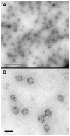
Figure 1. Analysis by negative staining electron microscopy and immunogold labeling of the chimeric yellow fever virus/dengue virus (YFV/DENV) particles secreted in the cell supernatant.
(A): Numerous 50 to 60 nm spherical particles that had morphological characteristics of a typical flavivirus were observed by regular negative staining electron microscopy. (B): The specificity of these particles was confirmed by immunogold labeling with an anti-DENV E glycoprotein. Scale bars : 0.5 µm in A ; 100 nm in B.
Figure 2
Figure 2. Analysis, by scanning electron microscopy (SEM), of Vero cells grown on microcarrier beads and infected with the chimeric YFV/DENV.
(A): At low magnification and before infection (day 0), Vero cells completely covered the microcarrier surface. (B): On day 0, analysis of these cells at high magnification showed them to have a smooth surface with protrusions. (C and D): On day 2 post-infection, several cells presented numerous chimeric viral particles at their surface (arrows). Scale bars : 100 µm in A ; 1 µm in B and C; 0.2 µm in D.
Figure 3
Figure 3. Analysis, by transmission electron microcopy (TEM), of Vero cells grown on microcarrier beads and infected with the chimeric YFV/DENV.
(A and B): Ultrathin sections of the cells attached to the microcarriers showed the presence of numerous chimeric viral particles in cisterns (black arrows) linked to the rough endoplasmic reticulum. Some virus-induced smooth vesicles were observed close to rough ER cisternae containing chimeric YFV/DENV particles (white arrows). Scale bars: 0.2 µm in A and B.
Figure 4
Figure 4. Principle underlying correlative scanning electron microscopy-transmission electron microscopy (CSEMTEM).
(A): SEM acquisition, at low magnification, made it possible to locate a microcarrier of particular interest (white arrow), i.e. bearing a cell with chimeric viral particles at its surface. (B): Mapping of the whole carbon disc with the multiscan module was performed, to identify the domain of interest on the disk (white circle). (C and D): After resizing (white circle in C), the sample containing the microcarrier of interest was examined under a stereo microscope (D). (E): After inclusion in Epon resin, thin sections (500 nm) of the resized sample were cut with an ultramicrotome and stained with toluidine blue, making it possible to identify the microcarrier of interest by light microscopy. (F): Ultrathin sections (70 nm) of this block were then cut for analysis of the resized sample by TEM, making it possible to visualize cells at the surface of the selected microcarrier (inset in F). The microcarrier of interest (arrows in A, D, E and F) can be followed during all these steps, through the recording of SEM, optical and TEM photographs. Scale bars : 200 µm in A, D and E; 2 mm in B and C; 100 µm in F.
Figure 5
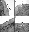
Figure 5. TEM analysis of Vero cells selected by the CSEMTEM method.
(A): Electron-dense chimeric viral particles were observed at the cell surface (black arrows). (B, C and D): Several released particles were observed at the site of small-vesicle exocytosis at the plasma membrane (white arrows). Scale bars: 0.2 µm in A, B and C; 0.1 µm in D.

References

    1. Murray L, Jones CT, Rice CM (2008) Architects of assembly: role of Flaviviridae non-structural proteins in virion morphogenesis. Nat Rev Microbiol 6: 699–708. - PMC - PubMed
    1. Shepard CW, Finelli L, Alter MJ (2005) Global epidemiology of hepatitis C virus infection. Lancet Infect Dis 5: 558–567. - PubMed
    1. Bhatt S, Gething PW, Brady OJ, Messina JP, Farlow AW, et al. (2013) The global distribution and burden of dengue. Nature 496: 504–507. - PMC - PubMed
    1. Gray EW, Nettleton PF (1987) The ultrastructure of cell cultures infected with border disease and bovine virus diarrhea viruses. J Gen Virol 68: 2239–2246. - PubMed
    1. Westaway EG, McKenzie JM, Kenney MT, Jones MK, Khromykh AA (1997) Ultrastructure of Kunjin virus-infected cells: colocalization of NS1 and NS3 with double-stranded RNA, and of NS2B with NS3, in virus-induced membrane structures. J Virol 71: 6650–6661. - PMC - PubMed

Publication types

LinkOut - more resources