. 2014 Apr;20(4):350-9.
doi: 10.1038/nm.3490. Epub 2014 Mar 30.

Kindlin-1 controls Wnt and TGF-β availability to regulate cutaneous stem cell proliferation

Affiliations

Kindlin-1 controls Wnt and TGF-β availability to regulate cutaneous stem cell proliferation

Emanuel Rognoni et al. Nat Med. 2014 Apr.

Abstract

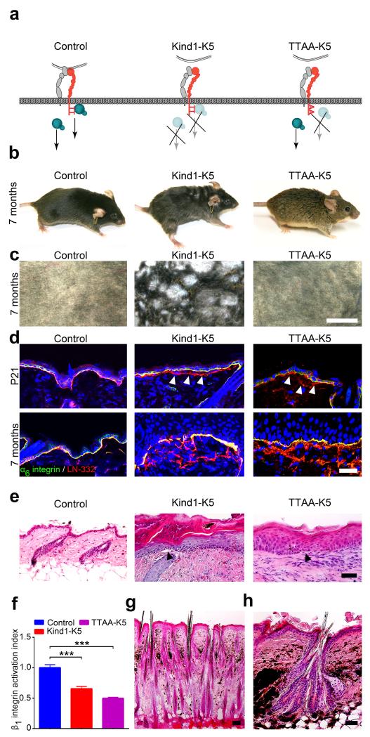
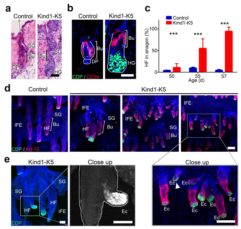
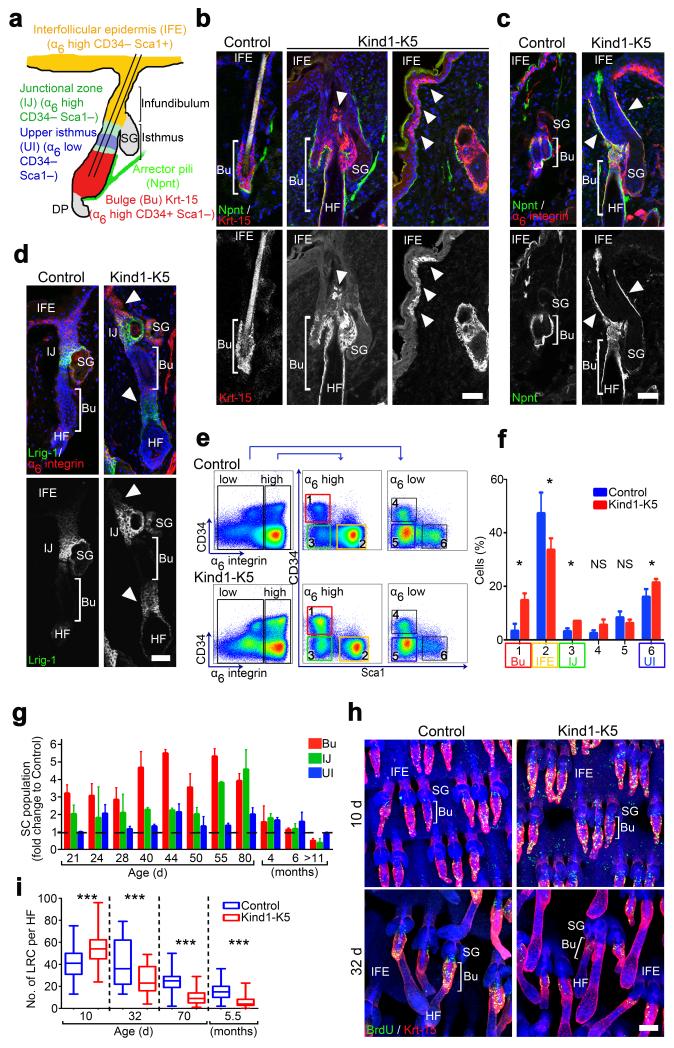
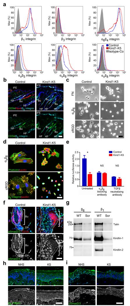
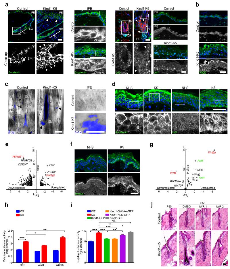
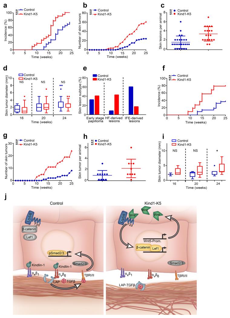
Kindlin-1 is an integrin tail binding protein that controls integrin activation. Mutations in the FERMT-1 gene, which encodes for Kindlin-1, lead to Kindler syndrome in man, which is characterized by skin blistering, premature skin aging and skin cancer of unknown etiology. Here we show that loss of Kindlin-1 in mouse keratinocytes recapitulates Kindler syndrome and also produces enlarged and hyperactive stem cell compartments, which lead to hyperthickened epidermis, ectopic hair follicle development and increased skin tumor susceptibility. Mechanistically, Kindlin-1 controls keratinocyte adhesion through β1-class integrins and proliferation and differentiation of cutaneous epithelial stem cells by promoting α(v)β(6) integrin-mediated transforming growth factor-β (TGF-β) activation and inhibiting Wnt-β-catenin signaling through integrin-independent regulation of Wnt ligand expression. Our findings assign Kindlin-1 the previously unknown and essential task of controlling cutaneous epithelial stem cell homeostasis by balancing TGF-β-mediated growth-inhibitory signals and Wnt-β-catenin-mediated growth-promoting signals.


Figures

Figure 1
Figure 1
Kindlin-1 controls HF growth. (a) Model of Kindlin-1 and β1 integrin functions in mice; Kind1-K5 mice lack Kindlin-1 in keratinocytes and TTAA-K5 mice express a Kindlin-binding deficient β1 integrin in which threonines-788/789 are substituted with alanines. (b) Appearance of 7 months old control, Kind1-K5 and TTAA-K5 mice. (c) View of shaved back skin of 7 months old control, Kind1-K5 and TTAA-K5 mice. (d) Immunofluorescence staining of back skin from P21 and 7 months old mice for LN-332 (red) and α6 integrin (green). Arrowheads indicate BM splitting. Nuclei are stained with DAPI (blue). (e) H/E staining of back skin from 6 months old mice. Kind1-K5 and TTAA-K5 skin shows hyperthickened skin, hyperkeratosis and blisters (arrowhead) at the epidermal-dermal junction. (f) Integrin activation index on primary control, Kind1-K5 and TTAA-K5 keratinocytes reported as mean 9EG7 binding normalized to total β1 integrin level ± SD (n=3 technical replicates). (g,h) H/E stained back skin sections of 7 months old Kind1-K5 mice. Scale bar indicates 5 mm in (c), 50 μm in (d,e,g) and 100 μm in (h).
Figure 2
Figure 2
Premature anagen induction and ectopic HF development in Kind1-K5 skin. (a) H/E stained back skin sections of P56 mice. (b) Immunostaining of HFs from P56 mice for CD34 (red) and CDP (green). Nuclei are stained with DAPI (blue). (c) The percentage of anagen HFs at indicated time points (mean ± SD, n=4 mice per genotype, ≥8 10× objective fields were counted). (d) Immunostaining of tail epidermal whole mounts for CDP (green), Krt-15 (red) and DAPI (blue) of P80 mice. Note ectopic HF growth in IFE of Kind1-K5 mice. (e) Ectopic HF outgrowth from a preexisting HF of the tail whole mount skin in a 6 months old Kind1-K5 mouse stained for CDP (green) and DAPI (blue). Scale bar indicates 50 μm in (a,b) and 100 μm in (d,e). Bu, bulge; SG, sebaceous gland; HG, hair germ; HF, hair follicle; DP, dermal papilla; IFE, interfollicular epidermis; Ec, ectopic HF.
Figure 3
Figure 3
Kindlin-1 regulates cutaneous epithelial SC homeostasis. (a) Distribution and expression of marker genes in SC subpopulations of murine HFs. (bd) Back skin immunostaining from 6 months old mice for Krt-15 (red) and Npnt (green) (b), Npnt (green) and α6 integrin (red) (c), and for Lrig1 (green) and α6 integrin (red) (d). Arrowheads indicate Krt-15 positive cells (b), Npnt deposition (c) and Lrig1 expression (d) in Kind1-K5 mice. (e) Keratinocytes from P40 old mice were separated by FACS into α6 integrin-high and -low and further analyzed for CD34 and Sca1 expression. (f) Gated populations (1–6) were quantified and represented as mean ± SD (n=5 Control and 3 Kind1-K5 mice). Color code in (e–f) corresponds to the cell population denoted in (a). (g) The relative amounts of SC subpopulations over time analyzed by FACS. Data show average fold increase (± SD) relative to control mice (n and P-values are listed in Supplementary Table 1). (h) BrdU (green) retaining cells in Krt-15 (red) stained tail whole mounts after indicated chase times. (i) Numbers of LRC per HF after indicated chasing periods were quantified as boxplots (10 d n=5 Control and 3 Kind1-K5; 32 d n=3 Control and 4 Kind1-K5; 80 d n=4 Control and 3 Kind1-K5). Boxplot whisker ends show Min/Max distribution and middle line reports the median. Nuclei are stained with DAPI (blue) (bd,h). Scale bar indicates 50 μm (bd) and 100 μm in (h). Bu, bulge; SG, sebaceous gland; HF, hair follicle; IFE, interfollicular epidermis; UI, upper isthmus; IJ, infundibulum junctional zone.
Figure 4
Figure 4
Kindlin-1 promotes αvβ6 integrin induced TGFβ release in vitro and in vivo. (a) Representative histogram of integrin levels on freshly isolated keratinocytes analyzed by FACS (n=3 biological replicates). (b) Immunofluorescence analysis of back skin from P44 mice for β6 integrin (cyan), CD34 (green) and Npnt (red). (c) Brightfield images of keratinocytes plated on FN-, αvβ6- and cRGD-coated surfaces. (d) Immunostaining of keratinocytes plated on αvβ6 and cRGD surfaces for paxillin (red) and F-actin or β6 integrin (green). (e) TGFβ release from extracellular matrix by keratinocytes from control or Kind1-K5 mice in the absence or presence of indicated blocking antibodies monitored with the tMLEC reporter cell line. Luciferase activity is reported as mean ± SEM (n=3 biological replicates). (f) Immunostaining of a HF from 4 months back skin for pSmad2/3 (green) and CD34 (red). (g) Streptavidin-bead pull-down assay with biotinylated wild type (WT) integrin tails or scrambled (Scr) peptides from keratinocyte lysates (representative blot from three independent experiments). (h,i) Immunofluorescence staining of skin sections from control and individuals with KS for β6 integrin (h) and pSmad2/3 (i). Nuclei are stained with DAPI (blue) (b,d,f,h,i). Scale bar indicates 50 μm (b,c,h,i) and 25 μm (d,f). NHS, normal human skin; KS, Kindler syndrome; Bu, bulge; DP, dermal papilla; HF, hair follicle; IFE, interfollicular epidermis.
Figure 5
Figure 5
Kindlin-1 controls Wnt-β-catenin signaling. (a) Immunofluorescence staining for β-catenin (green) of HF (P44) (left panel) and IFE (P55) (middle panel) and for Lef1 and CD34 (red) of HF and IFE (4 months) (right panel) from control and Kind1-K5 mice. Arrowheads indicate aberrant β-catenin (left panel) and Lef1 (right panel) localization. (b) IFE immunofluorescence staining for NICD (green) from P44 control and Kind1-K5 mice. (c) TOPgal reporter activity in tail HFs (left panel) and IFE (right panel) from 3 months old control and Kind1-K5 mice. Arrowheads indicate HF with abnormal TOPgal activity. (d) Immunofluorescence staining of human skin from normal and individuals with KS for β-catenin (left panel) and LEF1 (right panel). (e) Skin gene expression profile of NHS (n=3) versus KS (n=3) assessed with microarray and shown as volcano plot. Genes with ≥2 fold change in KS were plotted according to the Log2 fold change (x-axis) and Log10 P-value (y-axis). (f) Immunofluorescence staining for WNT5A (green) in skin of normal and individuals with KS. (g) Volcano plot of qPCR gene expression profile of keratinocytes from control versus Kind1-K5 mice. Mean expression relative to Gapdh of Wnt ligands (circle, red) and receptors (triangle, green) were plotted according to the Log2 fold change (x-axis) and log10 P-value (y-axis). (n=3 biological replicates, for mean ± SEM values see Supplementary Table 3). (h) Transient overexpression of GFP, Wnt4 and Wnt5a in floxed (WT) and Adeno-Cre treated Kindlin-1 deficient (KO) keratinocytes expressing the TOPFlash reporter. Values are corrected for the renilla control and represented as mean ± SEM fold increase relative to WT cells (n=5 biological replicates). (i) TOPFlash reporter activity in cells stably re-expressing Kindlin-1-GFP, integrin-binding deficient Kindlin-1-GFP, NLS-tagged Kindlin-1-GFP and Kindlin-2-GFP, respectively. Values are corrected for the renilla control, represented as fold increase relative to WT cells and reported as mean ± SEM (n=21 WT, KO; 13 Kind1-GFP; 9 Kind1-QW/AA-GFP, 5 Kind1-NLS-GFP, Kind2-GFP; all biological replicates). (j) H/E staining of control and Kind1-K5 mice one day (P50) and after treatment (P56) with indicated Wnt inhibitor. Nuclei are stained with DAPI (blue) (a,b,d,f). All scale bars indicate 50 μm. NHS, normal human skin; KS, Kindler syndrome, Bu, bulge; SG, sebaceous gland; HG, hair germ; HF, hair follicle; DP, dermal papilla; HB, hair bulb.
Figure 6
Figure 6
Loss of Kindlin-1 increases skin tumor incidence. (ae) Two stage carcinogenesis (n=26 Control and 23 Kind1-K5 mice). (a) Tumor incidence (P-value by log-rank test), (b) burden and (c) skin lesions per animal (reported as mean ± SD) after 25 weeks of treatment. (d) Tumor growth reported as diameter is shown in a boxplot where whisker ends are at 1.5 interquartile ranges and middle lines represent the median. (e) The percentage of skin lesion subtypes from control (n=25 lesions) and Kind1-K5 mice (n=47 lesions) were staged by histology and immunofluorescence analysis (see Supplementary Fig. 8a). (fi) One stage carcinogenesis with DMBA. (f) Tumor incidence, (g) skin lesion number, (h) frequency and (i) size of control (n=10) and Kind1-K5 (n=10) mice monitored as in (ad). (j) Molecular functions of Kindlin-1; in normal cells (left panel) Kindlin-1 activates β1-class integrins and αvβ6 integrin to facilitate adhesion and TGFβ liberation from LAP, respectively. Free TGFβ activates TGFβ receptors (TβRI/II) leading to nuclear translocation of phosphorylated Smad2/3, which promotes SC quiescence. In Kindlin-1 deficient cells (right panel) activation of β1-class integrins and αvβ6 is impaired leading to adhesion defects and loss of TGFβ-mediated SC quiescence. In addition, dysregulated Wnt ligand expression leading to elevation of Wnt5a leads to canonical Wnt-β-catenin signaling via the Lrp5/6-Fzd4 complex. Wnt5-Prom, Wnt5 promotor.

Comment in

  • Kindler syndrome in mice and men.
    Duperret EK, Ridky TW. Duperret EK, et al. Cancer Biol Ther. 2014 Sep;15(9):1113-6. doi: 10.4161/cbt.29482. Epub 2014 Jun 11. Cancer Biol Ther. 2014. PMID: 24919121 Free PMC article.

References

    1. Meves A, Stremmel C, Gottschalk K, Fässler R. The Kindlin protein family: new members to the club of focal adhesion proteins. Trends Cell Biol. 2009;19(10):504–513. - PubMed
    1. Lai-Cheong JE, et al. Kindler syndrome: a focal adhesion genodermatosis. Br J Dermatol. 2009;160(2):233–242. - PubMed
    1. Ussar S, et al. Loss of Kindlin-1 causes skin atrophy and lethal neonatal intestinal epithelial dysfunction. PLoS Genet. 2008;4(12):e1000289. - PMC - PubMed
    1. Montanez E, et al. Kindlin-2 controls bidirectional signaling of integrins. Genes Dev. 2008;22(10):1325–1330. - PMC - PubMed
    1. Moser M, Nieswandt B, Ussar S, Pozgajova M, Fässler R. Kindlin-3 is essential for integrin activation and platelet aggregation. Nat Med. 2008;14(3):325–330. - PubMed

References method section

    1. Fässler R, Meyer M. Consequences of lack of beta 1 integrin gene expression in mice. Genes Dev. 1995;9(15):1896–1908. - PubMed
    1. Ramirez A, et al. A keratin K5Cre transgenic line appropriate for tissue-specific or generalized Cre-mediated recombination. Genesis. 2004;39(1):52–57. - PubMed
    1. Abel EL, Angel JM, Kiguchi K, DiGiovanni J. Multi-stage chemical carcinogenesis in mouse skin: fundamentals and applications. Nat Protoc. 2009;4:1350–1362. - PMC - PubMed
    1. Kasper M, et al. Wounding enhances epidermal tumorigenesis by recruiting hair follicle keratinocytes. Proc Natl Acad Sci U S A. 2011;108:4099–4104. - PMC - PubMed
    1. Sundberg JP, Sundberg BA, Beamer WG. Comparison of chemical carcinogen skin tumor induction efficacy in inbred, mutant, and hybrid strains of mice: morphologic variations of induced tumors and absence of a papillomavirus cocarcinogen. Mol Carcinog. 1997;20:19–32. - PubMed

Publication types

MeSH terms

Supplementary concepts