Randomized Controlled Trial
. 2014 Jul;76(7):977-84.
doi: 10.1292/jvms.13-0338. Epub 2014 Mar 31.

Immunogenicity of a recombinant Mycobacterium smegmatis vaccine expressing the fusion protein CMX in cattle from Goiás State, Brazil

Affiliations
Randomized Controlled Trial

Immunogenicity of a recombinant Mycobacterium smegmatis vaccine expressing the fusion protein CMX in cattle from Goiás State, Brazil

Duanne Alves Da Silva et al. J Vet Med Sci. 2014 Jul.

Abstract

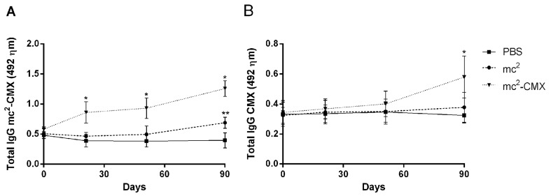
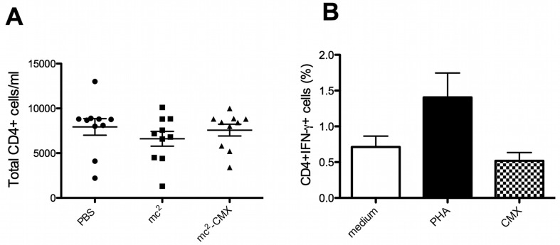
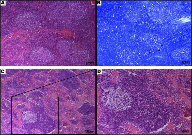
This study aimed to evaluate the immunogenicity of a recombinant Mycobacterium smegmatis vaccine expressing the CMX fusion protein composed of immunodominant epitopes Ag85C, MPT51 and HspX of Mycobacterium tuberculosis, which are important mycobacteria virulence factors. A group of Nelore heifers that were 10 to 12 months of age and negative for the tuberculin skin test (TST) were immunized with four doses of the recombinant vaccine mc(2)-CMX (M. smegmatis-Ag85C-MPT51-HspX) during a period of one year. Before each immunization, blood was collected to obtain sera for antibody analysis. Serological analysis demonstrated that mc(2)-CMX was able to induce a humoral response with increased levels of specific IgG antibodies against CMX, despite minimum antibody levels being detected for individual Ag85C, MPT51 or HspX recombinant antigens. However, there was no significant increase in specific CD4(+) IFN-γ-positive T cells. Lymphadenomegaly was observed in superficial cervical lymph nodes adjacent to the site of vaccination among mc(2)-CMX-vaccinated bovines, and the histopathological analysis demonstrated follicular hyperplasia without inflammatory infiltrate or granuloma formation. Animals remained negative for the TST until the end of the experiments, showing no cross-reactivity with the recombinant vaccine and tuberculin proteins. We discuss the potential of mc(2)-CMX to induce an immune response in cattle.


Figures

Fig. 1.
Fig. 1.
Immune humoral response induced by the recombinant vaccine. (A) Serum levels of anti-mc2-CMX antibodies among bovine immunized with mc2-CMX, mc2 and PBS. (B) Serum levels of anti-rCMX antibodies among bovine immunized with mc2CMX, mc2 and PBS. Animals were immunized with 1 × 10⁷ CFU/ml of each vaccine subcutaneously. Sera were obtained at different time points following vaccination and analyzed by ELISA. Each time point represents the mean and standard error of the mean of the optical density from 10 animals per group. * Statistically significant difference between the mc2-CMX and PBS groups (P<0.05). ** Statistically significant difference between the mc2 and PBS groups (P<0.05).
Fig. 2.
Fig. 2.
Induction of IFN-γ by CD4+ T cells. Cattle were revaccinated 200 days after the third immunization, and PBMCs were obtained. (A) Total CD4+ T cells from the vaccinated group were quantified by flow cytometry. The dots represent the number of CD4+ T cell/ml from each animal. The horizontal lines represent the average and standard deviation. (B) PBMCs from mc2-CMX-vaccinated animals were stimulated in vitro with medium, PHA or CMX, and the percentage of CD4+IFN-γ-positive T cells was analyzed. The bars represent the average and standard deviation per group. No significant statistical difference was observed.
Fig. 3.
Fig. 3.
Histopathological analysis of a superficial cervical lymph node from the mc2-CMX vaccinated group. The lymph node from a representative animal was sectioned and stained with hematoxylin eosin (H&E) (A, C and D) and Fite’s acid fast stain (B) 90 days after the last immunization of the group. Note the absence of inflammatory infiltrate or granuloma formation or necrosis (A). Fite’s acid fast staining revealed no presence of acid-fast bacilli (B). The structure of a normal lymph node with a germinal center, medular region and blood vessels can be seen (C and D).

Similar articles

Cited by

References

    1. Abadie V., Badell E., Douillard P., Ensergueix D., Leenen P. J. M., Tanguy M., Fiette L., Saeland S., Gicquel B., Winter N.2005. Neutrophils rapidly migrate via lymphatics after Mycobacterium bovis BCG intradermal vaccination and shuttle live bacilli to the draining lymph nodes. Blood 106: 1843–1850. doi: 10.1182/blood-2005-03-1281 - DOI - PubMed
    1. Ameni G., Vordermeier M., Aseffa A., Young D. B., Hewinson R. G.2010. Field evaluation of the efficacy of Mycobacterium bovis bacillus Calmette–Guerin against bovine tuberculosis in neonatal calves in Ethiopia. Clin. Vaccine Immunol. 17: 1533–1538. doi: 10.1128/CVI.00222-10 - DOI - PMC - PubMed
    1. Andersen P., Doherty T. M.2005. The success and failure of BCG — implications for a novel tuberculosis vaccine. Nat. Rev. Microbiol. 3: 656–662. doi: 10.1038/nrmicro1211 - DOI - PubMed
    1. Backus K. M., Boshoff H. I., Barry C. S., Boutureira O., Patel M. K.2011. Uptake of unnatural trehalose analogs as a reporter for Mycobacterium tuberculosis. Nat. Chem. Biol. 7: 228–235. doi: 10.1038/nchembio.539 - DOI - PMC - PubMed
    1. Belisle J. T., Vissa V. D., Sievert T., Takayama K., Brennan P. J., Besra G. S.1997. Role of the major antigen of Mycobacterium tuberculosis in cell wall biogenesis. Science 276: 1420–1422. doi: 10.1126/science.276.5317.1420 - DOI - PubMed

Publication types

MeSH terms