. 2014 Mar;26(3):1267-79.
doi: 10.1105/tpc.114.124495. Epub 2014 Mar 28.

A cyanobacterial chlorophyll synthase-HliD complex associates with the Ycf39 protein and the YidC/Alb3 insertase

Affiliations

A cyanobacterial chlorophyll synthase-HliD complex associates with the Ycf39 protein and the YidC/Alb3 insertase

Jack W Chidgey et al. Plant Cell. 2014 Mar.

Abstract

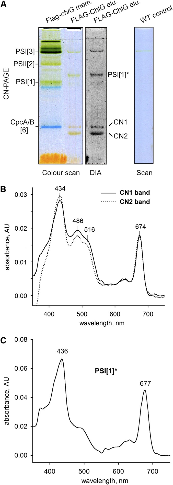
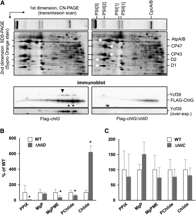
Macromolecular membrane assemblies of chlorophyll-protein complexes efficiently harvest and trap light energy for photosynthesis. To investigate the delivery of chlorophylls to the newly synthesized photosystem apoproteins, a terminal enzyme of chlorophyll biosynthesis, chlorophyll synthase (ChlG), was tagged in the cyanobacterium Synechocystis PCC 6803 (Synechocystis) and used as bait in pull-down experiments. We retrieved an enzymatically active complex comprising ChlG and the high-light-inducible protein HliD, which associates with the Ycf39 protein, a putative assembly factor for photosystem II, and with the YidC/Alb3 insertase. 2D electrophoresis and immunoblotting also provided evidence for the presence of SecY and ribosome subunits. The isolated complex contained chlorophyll, chlorophyllide, and carotenoid pigments. Deletion of hliD elevated the level of the ChlG substrate, chlorophyllide, more than 6-fold; HliD is apparently required for assembly of FLAG-ChlG into larger complexes with other proteins such as Ycf39. These data reveal a link between chlorophyll biosynthesis and the Sec/YidC-dependent cotranslational insertion of nascent photosystem polypeptides into membranes. We expect that this close physical linkage coordinates the arrival of pigments and nascent apoproteins to produce photosynthetic pigment-protein complexes with minimal risk of accumulating phototoxic unbound chlorophylls.


Figures

Figure 1.
Figure 1.
Purification of FLAG-ChlG and YidC-FLAG from Synechocystis Cells and Identification of Interacting Protein Partners. (A) FLAG-ChlG was purified from the Flag-chlG strain under native conditions on the anti-FLAG affinity gel. Eluted proteins were separated by SDS-PAGE together with a control pull-down from the Synechocystis wild type and stained with Coomassie blue. The amount of protein loaded for each sample corresponded to one-tenth of the total eluate volume. Designated protein bands were identified by MS (see Supplemental Table 1 for detected peptides). (B) Eluted proteins from the FLAG-ChlG pull-down were resolved by SDS-PAGE, transferred by immunoblot to a PVDF membrane and probed with selected antibodies. (C) Immunoprecipitation of the Ycf39 protein by the anti-Ycf39 antibody from wild-type and Flag-ChlG strains. The FLAG-ChlG protein that coimmunoprecipitated with Ycf39 was detected by an anti-FLAG antibody. (D) YidC-FLAG was purified from the yidC-Flag/ΔyidC strain and analyzed as described for FLAG-ChlG. (E) Eluted proteins from the YidC-FLAG pull-down were resolved by SDS-PAGE, blotted, and probed with the anti-ChlG antibody.
Figure 2.
Figure 2.
Separation of the Purified FLAG-ChlG Complex by CN-PAGE and Spectroscopy Analysis of Pigmented Bands. (A) CN-PAGE of the purified FLAG-ChlG complex and a control eluate from wild-type cells, the loading of which corresponded to ∼75% of the total eluates. Solubilized membranes (3 μg of chlorophyll) from the Flag-chlG strain were used to demonstrate the mobility of photosynthetic complexes: PSI[1] and PSI[3], monomer and trimer of PSI, respectively; PSII[2], dimer of PSII; CpcA/B[6], 107-kD heterohexamer of CpcA and CpcB phycobiliproteins. After separation, the gel was scanned in color by an office scanner and in transmittance mode (DIA) using a LAS 4000 imager (Fuji). CN1, CN2, and PSI[1]* mark protein complexes identified in FLAG-ChlG elution (line 2) but not in the total membrane fraction (line 1). (B) The orange-yellow CN1 and CN2 bands were excised from the CN-PAGE gel, and absorption spectra were recorded as described in Methods. AU, absorbance units. (C) Absorption spectra of the PSI[1]* complex; the red shifted absorbance of the chlorophyll Qy peak is typical for PSI complexes.
Figure 3.
Figure 3.
2D Electrophoresis of the Purified FLAG-ChlG Complex and Identification of Individual Protein Spots. A gel strip from CN-PAGE with separated FLAG-ChlG complexes (see Figure 2A) was further separated in a second dimension by SDS-PAGE. The gel was stained by Sypro Orange and then blotted onto a PVDF membrane. The identity of designated spots on the stained gel was assigned by MS and further verified by specific antibodies (see Results). PSI[1]* is a form of monomeric PSI migrating slower than a typical PSI[1] (Figures 2A and 2D). YidC, SecY, and Ycf39 were detected using specific antibodies. For identification of individual ribosome subunits migrating close to the top of CN gel, see Supplemental Figure 5.
Figure 4.
Figure 4.
Separation of the Purified FLAG-ChlG Complexes by Gel Filtration Chromatography and Immunodetection of Eluted Proteins. The FLAG-ChlG pull-down was loaded on a BioSec 3000 column, and eluted protein/complexes were detected by absorbance at 280 and 440 nm. Eluted fractions were collected and subjected to immunoblot analysis; each band corresponds to the 0.2-mL chromatograph fraction with which it is aligned. DDM, dodecyl-β-maltoside.
Figure 5.
Figure 5.
Analysis of Pigments Associated with the FLAG-ChlG-HliD Complex. (A) FLAG-ChlG pull-down obtained from the PSI-less background was separated on a gel filtration column essentially as described for Figure 4 and the GF3 peak collected (gray box). DDM, dodecyl-β-maltoside. (B) Absorption spectra of the GF3 and GF4 peaks were recorded by a HPLC diode array detector. (C) Pigments were extracted from the pooled fractions representing the GF3 peak (gray box in [A]), separated by HPLC and detected at 415 nm. The area of each peak was integrated and the molar stoichiometry of individual pigments estimated. Myxo, myxoxanthophyll; Zea, zeaxanthin; GG-Chl, geranylgeranyl chlorophyll; β-Car, β-carotene.
Figure 6.
Figure 6.
ChlG Activity of the Purified FLAG-ChlG Complex. (A) Conversion of the endogenous Chlide pool and exogenous Chlide to geranylgeranyl-chlorophyll (GG-Chl). Stopped assays were performed in the presence of 20 μM geranylgeranyl diphosphate. Assays were performed in triplicate and each time point was analyzed by reverse-phase HPLC. (B) Utilization of exogenously added Chlide by FLAG-ChlG. Assays were performed as in (A) but with the addition of 20 μM Chlide. Each chromatography trace is representative of one of three replicates. (C) Evolution of the geranylgeranyl-chlorophyll product in the exogenous Chlide assays in (B).
Figure 7.
Figure 7.
Characterization of Synechocystis ΔhliD Strains. (A) Deletion of the hliD gene affects abundance of FLAG-ChlG and Ycf39 proteins as well as the formation of higher mass FLAG-ChlG complexes in the membrane. Membrane fractions isolated from the Synechocystis Flag-chlG (left) and Flag-chlGhliD (right) strains were separated by 2D CN/SDS-PAGE, stained by Sypro Orange, and finally blotted onto a PVDF membrane. Only the part of the SDS gel between ∼25 and ∼70 kD is presented; spots representing PSII core subunits and AtpA/B proteins are marked by white arrows. FLAG-ChlG and Ycf39 were detected by anti-FLAG and anti-Ycf39 antibodies, respectively. Black asterisks indicate the positions of the CN1 and CN2 bands (see Figure 3); the black arrowhead indicates a putative complex between FLAG-ChlG and Ycf39. In the lowest immunoblot panel, the Ycf39 signal was overexposed. (B) Analysis of chlorophyll precursors in the wild-type and ΔhliD strains grown photoautotrophically at 40 μmol of photons m−2 s−1. Precursors were extracted with 70% methanol from cells at OD750 = 0.4 and analyzed by HPLC equipped with a diode array detector and a pair of fluorescence detectors (Hollingshead et al., 2012). PPIX, protoporphyrin IX; MgP, Mg-protoporphyrin IX; MgPME, Mg-protoporphyrin IX monomethylester; PChlide, protochlorophyllide. Values shown represent means ± sd from three independent measurements. Asterisks indicate statistically significant differences in precursor levels as tested using a paired t test (P = 0.05). (C) An identical measurement of chlorophyll precursors as in (B) performed on the ΔhliC strain.
Figure 8.
Figure 8.
Immunodetection of HliD and ChlG in the Synechocystis Δycf39 Strain. Membrane fractions prepared from wild-type and Δycf39 strains were separated by SDS-PAGE and blotted, and the HliD and ChlG proteins were detected by specific antibodies. The level of cytochrome f detected though its peroxidase activity was included as a loading control.

Similar articles

Cited by

References

    1. Addlesee H.A., Fiedor L., Hunter C.N. (2000). Physical mapping of bchG, orf427, and orf177 in the photosynthesis gene cluster of Rhodobacter sphaeroides: Functional assignment of the bacteriochlorophyll synthetase gene. J. Bacteriol. 182: 3175–3182. - PMC - PubMed
    1. Antonoaea R., Fürst M., Nishiyama K., Müller M. (2008). The periplasmic chaperone PpiD interacts with secretory proteins exiting from the SecYEG translocon. Biochemistry 47: 5649–5656. - PubMed
    1. Apel K., Hirt H. (2004). Reactive oxygen species: Metabolism, oxidative stress, and signal transduction. Annu. Rev. Plant Biol. 55: 373–399. - PubMed
    1. Beck K., Eisner G., Trescher D., Dalbey R.E., Brunner J., Müller M. (2001). YidC, an assembly site for polytopic Escherichia coli membrane proteins located in immediate proximity to the SecYE translocon and lipids. EMBO Rep. 2: 709–714. - PMC - PubMed
    1. Bhaya D., Dufresne A., Vaulot D., Grossman A. (2002). Analysis of the hli gene family in marine and freshwater cyanobacteria. FEMS Microbiol. Lett. 215: 209–219. - PubMed

Publication types

MeSH terms

LinkOut - more resources