. 2014 Apr 15;5(7):1869-84.
doi: 10.18632/oncotarget.1853.

The immunocytokine NHS-IL12 as a potential cancer therapeutic

Affiliations

The immunocytokine NHS-IL12 as a potential cancer therapeutic

Jonathan Fallon et al. Oncotarget. .

Abstract

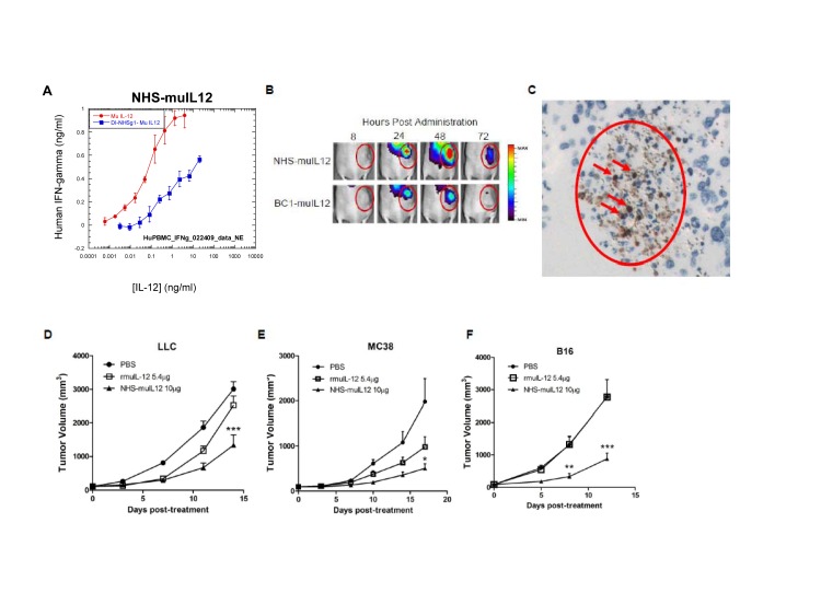
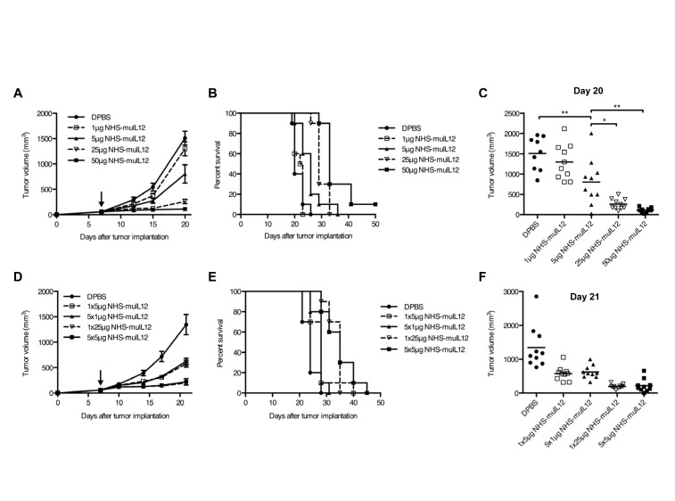
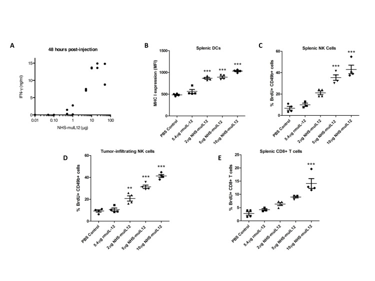
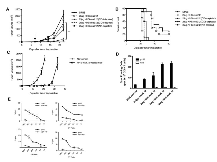
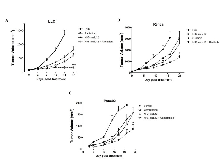
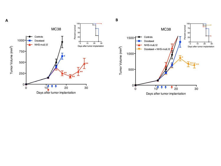
Targeted delivery of IL-12 might turn this cytokine into a safer, more effective cancer therapeutic. Here we describe a novel immunocytokine, NHS-IL12, consisting of two molecules of IL-12 fused to a tumor necrosis-targeting human IgG1 (NHS76). The addition of the human IgG1 moiety resulted in a longer plasma half-life of NHS-IL12 than recombinant IL-12, and a selective targeting to murine tumors in vivo. Data from both in vitro assays using human PBMCs and in vivo primate studies showed that IFN-gamma production by immune cells is attenuated following treatment with the immunocytokine, suggesting an improved toxicity profile than seen with recombinant IL-12 alone. NHS-IL12 was superior to recombinant IL-12 when evaluated as an anti-tumor agent in three murine tumor models. Mechanistic studies utilizing immune cell subset-depleting antibodies, flow cytometric methods, and in vitro cytotoxicity and ELISA assays all indicated that the anti-tumor effects of NHS-IL12 were primarily CD8+ T cell-dependent and likely IL-12-mediated. Combining NHS-IL12 treatment with a cancer vaccine, radiation, or chemotherapy resulted in greater anti-tumor effects than each individual therapy alone. These preclinical findings provide a rationale for the clinical testing of this immunocytokine, both as a single agent and in combination with vaccines, radiation and chemotherapy.


Conflict of interest statement

RT, GK, WG, AB, BN, YL, and HS are employees of EMD Serono. The other authors do not have any potential conflicts of interest to disclose.

Figures

Figure 1
Figure 1. Conjugation of human IL-12 to a DNA/histone-binding antibody increases its half-life and attenuates its ability to stimulate IFN-gamma production
(A) Ribbon diagram of NHS-IL12, an immunocytokine consisting of the NHS76 antibody fused to 2 IL-12p70 heterodimers. (B) Human PBMCs were stimulated in vitro with 2 μg/ml PHA for 4 days, and then an additional day following the addition of 10ng/ml rHuIL-2. NHS-huIL12 (blue) or rHuIL-12 (red) was then added to the cells, and supernatant samples were collected 24 h later. Human IFN-gamma levels in the supernatant were determined by ELISA. Results show mean ± SE of triplicate wells for each condition. (C) Cynomolgus macaques were treated with 40 μg/kg NHS-huIL12 s.c. (green), 40 μg/kg NHS-huIL12 intravenously (i.v., pink), or 4 μg/kg rHuIL-12 i.v. (red). Serum IFN-gamma levels in 2 animals per group were monitored for 5 days after injection. (D-F) Pharmacokinetic profile of NHS-huIL12 compared to rHuIL-12 in cynomolgus macaques. Cynomolgus macaques were treated with (D) 40 μg/kg NHS-huIL12 s.c., (E) 40 μg/kg NHS-huIL12 intravenously (i.v.), or (F) 4 μg/kg rHuIL-12 i.v. Plasma drug levels in 2 animals per treatment group were monitored for 8 days following injection.
Figure 2
Figure 2. Fusion of murine IL-12 to NHS76 promotes its uptake by tumors and improves its anti-tumor efficacy
(A) Human PBMCs were stimulated in vitro with 2 μg/ml PHA for 4 days and then an additional day in the presence of 10ng/ml rHuIL-2. NHS-muIL12 (blue) or rMuIL-12 (red) were then added to the cells, and supernatant samples were collected 24 h later. Human IFN-gamma levels in the supernatant were determined by ELISA. Results show mean ± SE of triplicate wells for each condition. (B) Athymic mice bearing subcutaneous LLC tumors were injected with 100 μg of fluorescence-conjugated NHS-muIL12 or BC1-muIL12 and imaged at 8, 24, 48, and 72 h post-injection. One representative mouse from each treatment group is shown. (C) LLC tumor sections revealed that NHS-muIL12 binds to exposed cell nuclei within necrotic regions of murine tumors. (D-F) NHS-muIL12 displayed greater anti-tumor activity than an equimolar dose of rMuIL-12 against subcutaneous (D) LLC, (E) MC38, and (F) B16 tumors (n = 8 mice per group). Average tumor volumes ± SE are shown. Asterisks indicate statistically significant differences between the NHS-muIL12 treatment group versus the control or rMuIL-12 treatment groups (2-way ANOVA followed by Bonferroni's post-test; *, P < 0.05; **, P < 0.01; ***, P <0.001).
Figure 3
Figure 3. The anti-tumor activity of NHS-muIL12 is dose-dependent and is unaffected by dose fractionation
(A–C) Immunocompetent C57BL/6 mice bearing subcutaneous MC38 tumors were randomized (n = 9-10 mice per group) and treated with a single s.c. injection of DPBS (closed circle) or 1 μg (open square), 5 μg (closed triangle), 25 μg (open triangle), or 50 μg (closed square) NHS-muIL12. Graphs of (A) mean tumor volume, (B) overall survival, and (C) individual tumor volumes on day 20 are shown. Arrow in panel A indicates treatment on day 7, and error bars indicate SEM. Asterisks indicate statistically significant differences between mean tumor volumes on day 20 (1-way ANOVA with Tukey's post-test; *, P < 0.05; **, P < 0.01). (D–F) To determine whether dose fractionation affected the anti-tumor activity of NHS-muIL12, MC38 tumor-bearing mice were randomized (n = 10 mice per group) and treated with DPBS (closed circle) or 1x5 μg (open square), 5x1 μg (closed triangle), 1x25 μg (open triangle), or 5x5 μg (closed square) NHS-muIL12. Graphs of (D) mean tumor volume, (E) overall survival, and (F) individual tumor volumes on day 21 are shown. Arrow in panel D indicates treatment initiation on day 7, and error bars indicate SEM. A 1-way ANOVA followed by Tukey's multiple comparisons test were used to compare mean tumor volumes on day 21. The mean differences observed at this time point between each NHS-muIL12 treatment group relative to the control group were statistically significant (P < 0.0001).
Figure 4
Figure 4. Immune activation markers correlate with increasing doses of NHS-muIL12
(A) C57BL/6 mice were injected with a single dose of NHS-muIL12 within the range of 0.01–50 μg. For each dose level, 2 mice were injected and serum samples were obtained 48 h later for IFN-gamma detection by ELISA. (B–E) MC38 tumor-bearing mice were randomized (n = 4 mice per group) and treated with PBS (closed circle), 5.4 μg rMuIL-12 (closed square), 2 μg NHS-muIL12 (closed triangle), 5 μg NHS-muIL12 (closed inverted triangle), or 10 μg NHS-muIL12 (closed diamond). (B) Flow cytometry was used to quantify MHC class I expression levels on splenic dendritic cells 5 days after treatment. Percent BrdU incorporation by (C) splenic NK, (D) tumor-infiltrating NK, and (E) splenic CD8+ T cells was also evaluated 5 days after treatment by flow cytometry. Error bars indicate SEM. Asterisks indicate statistically significant differences between NHS-muIL12 treated groups versus control and rMuIL-12 treated groups (1-way ANOVA with Bonferroni's post-test; **, P < 0.01; ***, P <0.001).
Figure 5
Figure 5. The anti-tumor activity of NHS-muIL12 depends on CD8+ T cells
(A–C) MC38 tumor-bearing mice were randomized and treated with DPBS (closed circle), 25 μg NHS-muIL12 (open square), or 25 μg NHS-muIL12 together with antibodies to deplete CD4 (closed triangle), CD8 (open triangle), or NK cells (closed square). Arrow denotes NHS-muIL12 treatment on day 7. Graphs representing (A) mean tumor volume over time and (B) overall survival are shown. Differences in mean tumor volumes on day 22 between mice treated with 25 μg NHS-muIL12 alone and together with CD8+ cell-depleting antibodies were statistically significant (1-way ANOVA with Bonferroni's post-test, ***, P < 0.001). (C) Four mice from this study became tumor-free following NHS-muIL12 treatment, and were rechallenged 4 months later with 300,000 MC38 cells on the opposite rear flank (open square). Five naïve C57BL/6 mice were challenged with MC38 tumors in the same way (closed circle) (t-test; *, P < 0.05). Error bars in panels A and C indicate SEM. (D) MC38 tumor-bearing mice were randomized and treated with PBS, 5.4 μg rMuIL-12, 2 μg NHS-muIL12, 5 μg NHS-muIL12, or 10 μg NHS-muIL12. Five days after treatment, splenocytes from 4 mice per treatment group were pooled and stimulated for 6 days in vitro with 1 μg/ml p15E peptide. ELISPOT assays were then used to determine the relative frequencies of splenic CD8+ T cells specific for p15E or ovalbumin. (E) The spleens of 4 CEA.Tg mice that were completely cured of MC38/CEA+ tumors following NHS-muIL12 treatment were tested individually for the presence of a p15E-specific CTL response. Results shown in panels D and E are mean ± SE of triplicate wells.
Figure 6
Figure 6. The combination of NHS-muIL12 with an experimental cancer vaccine enhances the anti-tumor response
(A) Wild-type C57BL/6 and MUC1.Tg mice were treated with cyclophosphamide and then vaccinated 3 times with tecemotide. Isolated CD4+ splenocytes from vaccinated and naïve mice of each strain were stimulated in vitro with MUC1 BP25 peptide, and lymphoproliferation was measured by [3H]-thymidine incorporation. Results show mean proliferation (counts per minute) ± SE of triplicate wells for each condition. (B–C) MUC1.Tg mice bearing (B) MC38/MUC1+ s.c. tumors or (C) Panc02/MUC1+ orthotopic tumors were randomized and treated with control liposomes, cyclophosphamide and tecemotide, cyclophosphamide and NHS-muIL12, or the combination of cyclophosphamide, tecemotide, and NHS-muIL12 (n = 9-10 mice per group). Cyclophosphamide (2 mg i.v.) was administered on day 0. NHS-muIL12 (10 μg s.c.) was given on day 1. Tecemotide (4x25 μg per dose) was administered s.c. on days 1, 8, 15, and 22. Results show mean tumor volume ± SE over time for each treatment group. Differences in mean tumor volume between groups treated with cyclophosphamide, tecemotide, and NHS-muIL12 were statistically significant versus groups treated with cyclophosphamide and NHS-muIL12 or tecemotide (2-way ANOVA with Bonferonni's post-test; *, P < 0.05; ***, P <0.001).
Figure 7
Figure 7. The combination of NHS-muIL12 with fractionated radiotherapy or chemotherapy enhances the anti-tumor response
(A) Beginning on day 0, LLC tumor-bearing mice were randomized (n = 9 mice per group) and treated with PBS, fractionated radiotherapy alone (360 cGy on days 0-4), NHS-muIL12 alone (10 μg s.c. on day 0), or the combination of fractionated radiotherapy plus NHS-muIL12. (B) Beginning on day 0, Renca tumor-bearing Balb/c mice were randomized (n = 10 mice per group) and treated with PBS, sunitinib alone (20 mg/kg orally on days 0-6), NHS-muIL12 alone (10 μg s.c. on day 1), or sunitinib plus NHS-muIL12. (C) Panc02 tumor-bearing mice were randomized (n = 11 mice per group) and treated with PBS, gemcitabine alone (120mg/kg i.v. on days 0 and 14), NHS-muIL12 alone (10 μg s.c. on day 0), or gemcitabine plus NHS-muIL12. Results show mean tumor volume ± SE over time for each treatment group. Differences between groups treated in combination were statistically significant versus groups that received monotherapies (2-way ANOVA with Bonferonni's post-test; *, P < 0.05; ***, P <0.001).
Figure 8
Figure 8. Docetaxel and NHS-muIL12 are effective as monotherapies and combined therapies in mice bearing well-established MC38 tumors
(A) Mice bearing well-established (~150 mm3) s.c. MC38 tumors were randomized (n = 12 mice per group) and treated with saline (closed circle), 3x0.5 mg docetaxel (closed square) on days 11, 13 and 15 (blue arrows), or 1x50 μg NHS-muIL12 (closed triangle) on day 11 (red arrow) after tumor implantation. Docetaxel and NHS-muIL12 each significantly delayed tumor growth as compared with saline-treated controls (1-way ANOVA with Bonferroni's post-test; *, P < 0.05; **, P < 0.0001). (B) To study the combined efficacy of these therapeutic agents, mice bearing MC38 tumors were randomized (n = 12 mice per group) and treated with saline (closed circle), 3x0.5 mg docetaxel (closed square) on days 11, 13 and 15 (blue arrows), 1x50 μg NHS-muIL12 (closed triangle) on day 18 (red arrow), or the sequential combination of both (closed inverted triangle). Graphs show mean tumor volume ± SE over time for each treatment group. The sequential treatment combination resulted in significantly greater anti-tumor effects than saline-treated controls (1-way ANOVA with Tukey's post-test; *, P < 0.05; **, P < 0.0001). Inserts in both panels A and B indicate percent survival of each treatment group over time following tumor implantation.

References

    1. Trinchieri G. Interleukin-12 and the regulation of innate resistance and adaptive immunity. Nat Rev Immunol. 2003;3:133–46. - PubMed
    1. Manetti R, Parronchi P, Giudizi MG, Piccinni MP, Maggi E, Trinchieri G, Romagnani S. Natural killer cell stimulatory factor (Interleukin 12 [IL-12]) induces T helper type 1 (Th1)-specific immune responses and inhibits the development of IL-4-producing Th cells. J Exp Med. 1993;177:1199–204. - PMC - PubMed
    1. Gately MK, Desai BB, Wolitzky AG, Quinn PM, Dwyer CM, Podlaski FJ, Familletti PC, Sinigaglia F, Chizonnite R, Gubler U, Stern AS. Regulation of human lymphocyte proliferation by a heterodimeric cytokine, IL-12 (cytotoxic lymphocyte maturation factor) J Immunol. 1991;147:874–82. - PubMed
    1. Perussia B, Chan SH, D'Andrea A, Tsuji K, Santoli D, Pospisil M, Young D, Wolf SF, Trinchieri G. Natural killer (NK) cell stimulatory factor or IL-12 has differential effects on the proliferation of TCR-αβ+, TCR-γδ+ T lymphocytes, and NK cells. J Immunol. 1992;149:3495–502. - PubMed
    1. Wong HL, Wilson DE, Jenson JC, Familletti PC, Stremlo DL, Gately MK. Characterization of a factor(s) which synergizes with recombinant interleukin 2 in promoting allogeneic human cytolytic T-lymphocyte responses in vitro. Cell Immunol. 1988;111:39–54. - PubMed

Publication types

MeSH terms