

Review
doi: 10.1128/AEM.00699-14.
Epub 2014 Mar 28.
Raman spectroscopy of microbial pigments
Affiliations
- PMID: 24682303
- PMCID: PMC4018853
- DOI: 10.1128/AEM.00699-14
Item in Clipboard
Review
Raman spectroscopy of microbial pigments
Appl Environ Microbiol.
2014 Jun.
Abstract
Raman spectroscopy is a rapid nondestructive technique providing spectroscopic and structural information on both organic and inorganic molecular compounds. Extensive applications for the method in the characterization of pigments have been found. Due to the high sensitivity of Raman spectroscopy for the detection of chlorophylls, carotenoids, scytonemin, and a range of other pigments found in the microbial world, it is an excellent technique to monitor the presence of such pigments, both in pure cultures and in environmental samples. Miniaturized portable handheld instruments are available; these instruments can be used to detect pigments in microbiological samples of different types and origins under field conditions.
Figures
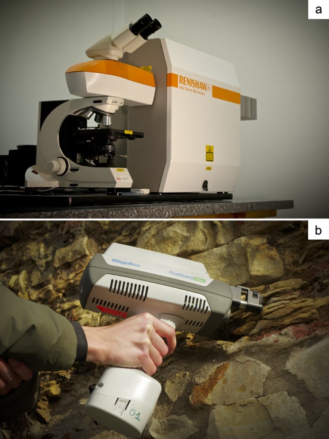
(a) Conventional confocal Raman microspectrometer. The laser irradiates the object selected in the optical microscope (laser spot ≈ 2 μm), the scattered light from the sample is collected by the optics of the microscope passing through holographic filters, pinhole, and monochromator to be detected by a charge-coupled device (CCD). (b) Handheld Raman spectrometer allows direct measurements on outcrops (cyanobacteria on siliciclastic rocks at Albertov, Prague, Czech Republic). The rugged probe head permits laser illumination of the object and collection of the scattered light.
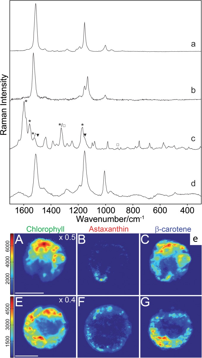
Examples of microbiological application of Raman spectroscopy using different instrumentation. (a to c) Raman spectra obtained using a laboratory Raman microspectrometer. Raman spectra of salinixanthin from a culture of Salinibacter ruber (a) (532-nm wavelength; image modified from reference 54), of flexirubin from a culture of Flavobacterium johnsonae (b) (532-nm wavelength; image modified from reference 57), and of chlorophyll (□), carotenoid (▼), and scytonemin (*) from a native gypsum crust (Yungay, Atacama, Chile) (c) (785-nm wavelength; image modified from reference 69). (d) Raman spectrum collected using a handheld Raman spectrometer of a spirilloxanthin-like carotenoid of the red layer of the bottom gypsum crust from a saltern at Eilat (Israel) colonized by Halochromatium-like microorganisms (532-nm wavelength; image modified from reference 117). (e) Example of Raman mapping–distribution of carotenoids (astaxanthin, β-carotene, chlorophyll in the frame of flagellated motile cell obtained using a confocal Raman spectrometer (532-nm wavelength; image modified from reference , which was published under a Creative Commons agreement). The intensities within the concentration map represent relative component concentrations. Bars, 10 μm.
Similar articles
-
Single-cell pigment analysis of phototrophic and phyllosphere bacteria using simultaneous detection of Raman and autofluorescence spectra.Appl Environ Microbiol. 2025 May 21;91(5):e0012925. doi: 10.1128/aem.00129-25. Epub 2025 Apr 10. Appl Environ Microbiol. 2025. PMID: 40207966 Free PMC article.
-
Assessment of Biotechnologically Important Filamentous Fungal Biomass by Fourier Transform Raman Spectroscopy.Int J Mol Sci. 2021 Jun 23;22(13):6710. doi: 10.3390/ijms22136710. Int J Mol Sci. 2021. PMID: 34201486 Free PMC article.
-
Characterization of carotenoids in soil bacteria and investigation of their photodegradation by UVA radiation via resonance Raman spectroscopy.Analyst. 2015 Jul 7;140(13):4584-93. doi: 10.1039/c5an00438a. Analyst. 2015. PMID: 26029748
-
Cultivation-Free Raman Spectroscopic Investigations of Bacteria.Trends Microbiol. 2017 May;25(5):413-424. doi: 10.1016/j.tim.2017.01.002. Epub 2017 Feb 7. Trends Microbiol. 2017. PMID: 28188076 Review.
-
Raman spectroscopy in halophile research.Front Microbiol. 2013 Dec 10;4:380. doi: 10.3389/fmicb.2013.00380. Front Microbiol. 2013. PMID: 24339823 Free PMC article. Review.
Cited by
-
Classification and identification of pigmented cocci bacteria relevant to the soil environment via Raman spectroscopy.Environ Sci Pollut Res Int. 2015 Dec;22(24):19317-25. doi: 10.1007/s11356-015-4593-5. Epub 2015 May 5. Environ Sci Pollut Res Int. 2015. PMID: 25940486
-
Biosignature stability in space enables their use for life detection on Mars.Sci Adv. 2022 Sep 9;8(36):eabn7412. doi: 10.1126/sciadv.abn7412. Epub 2022 Sep 7. Sci Adv. 2022. PMID: 36070383 Free PMC article.
-
Pilot SERS Monitoring Study of Two Natural Hypersaline Lake Waters from a Balneary Resort during Winter-Months Period.Biosensors (Basel). 2023 Dec 29;14(1):19. doi: 10.3390/bios14010019. Biosensors (Basel). 2023. PMID: 38248396 Free PMC article.
-
Raman Spectroscopy and Its Modifications Applied to Biological and Medical Research.Cells. 2022 Jan 24;11(3):386. doi: 10.3390/cells11030386. Cells. 2022. PMID: 35159196 Free PMC article. Review.
-
Effects of nicotine on the biosynthesis of carotenoids in halophilic Archaea (class Halobacteria): an HPLC and Raman spectroscopy study.Extremophiles. 2018 May;22(3):359-366. doi: 10.1007/s00792-018-0995-x. Epub 2018 Jan 15. Extremophiles. 2018. PMID: 29335805
References
-
- Oren A. 2002. Pigments of halophilic microorganisms, p 173–206 In Halophilic microorganisms and their environments: cellular origin, life in extreme habitats and astrobiology. Springer, Dordrecht, The Netherlands
-
- Koyama Y. 1991. Structures and functions of carotenoids in photosynthetic systems. J. Photochem. Photobiol. B 9:265–280. 10.1016/1011-1344(91)80165-E - DOI
Publication types
MeSH terms
Substances
LinkOut - more resources
Full Text Sources
Other Literature Sources
Medical
