

The chaperone domain BRICHOS prevents CNS toxicity of amyloid-β peptide in Drosophila melanogaster
- PMID: 24682783
- PMCID: PMC4036473
- DOI: 10.1242/dmm.014787
The chaperone domain BRICHOS prevents CNS toxicity of amyloid-β peptide in Drosophila melanogaster
Abstract
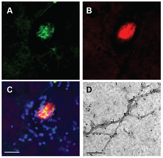
Aggregation of the amyloid-β peptide (Aβ) into toxic oligomers and amyloid fibrils is linked to the development of Alzheimer's disease (AD). Mutations of the BRICHOS chaperone domain are associated with amyloid disease and recent in vitro data show that BRICHOS efficiently delays Aβ42 oligomerization and fibril formation. We have generated transgenic Drosophila melanogaster flies that express the Aβ42 peptide and the BRICHOS domain in the central nervous system (CNS). Co-expression of Aβ42 and BRICHOS resulted in delayed Aβ42 aggregation and dramatic improvements of both lifespan and locomotor function compared with flies expressing Aβ42 alone. Moreover, BRICHOS increased the ratio of soluble:insoluble Aβ42 and bound to deposits of Aβ42 in the fly brain. Our results show that the BRICHOS domain efficiently reduces the neurotoxic effects of Aβ42, although significant Aβ42 aggregation is taking place. We propose that BRICHOS-based approaches should be explored with an aim towards the future prevention and treatment of AD.
Keywords: Alzheimer’s disease; Amyloid; Chaperone; Protein misfolding.
© 2014. Published by The Company of Biologists Ltd.
Figures


Similar articles
-
Dementia-related Bri2 BRICHOS is a versatile molecular chaperone that efficiently inhibits Aβ42 toxicity in Drosophila.Biochem J. 2016 Oct 15;473(20):3683-3704. doi: 10.1042/BCJ20160277. Epub 2016 Aug 11. Biochem J. 2016. PMID: 27514716
-
Amyloid Fibril Formation of Arctic Amyloid-β 1-42 Peptide is Efficiently Inhibited by the BRICHOS Domain.ACS Chem Biol. 2022 Aug 19;17(8):2201-2211. doi: 10.1021/acschembio.2c00344. Epub 2022 Jul 25. ACS Chem Biol. 2022. PMID: 35876740 Free PMC article.
-
Identification of potential aggregation hotspots on Aβ42 fibrils blocked by the anti-amyloid chaperone-like BRICHOS domain.Nat Commun. 2024 Feb 1;15(1):965. doi: 10.1038/s41467-024-45192-4. Nat Commun. 2024. PMID: 38302480 Free PMC article.
-
BRICHOS domain associated with lung fibrosis, dementia and cancer--a chaperone that prevents amyloid fibril formation?FEBS J. 2011 Oct;278(20):3893-904. doi: 10.1111/j.1742-4658.2011.08209.x. Epub 2011 Jul 5. FEBS J. 2011. PMID: 21668643 Review.
-
Transthyretin and BRICHOS: The Paradox of Amyloidogenic Proteins with Anti-Amyloidogenic Activity for Aβ in the Central Nervous System.Front Neurosci. 2017 Mar 15;11:119. doi: 10.3389/fnins.2017.00119. eCollection 2017. Front Neurosci. 2017. PMID: 28360830 Free PMC article. Review.
Cited by
-
On the role of sidechain size and charge in the aggregation of Aβ42 with familial mutations.Proc Natl Acad Sci U S A. 2018 Jun 26;115(26):E5849-E5858. doi: 10.1073/pnas.1803539115. Epub 2018 Jun 12. Proc Natl Acad Sci U S A. 2018. PMID: 29895690 Free PMC article.
-
Functionalization of amyloid fibrils via the Bri2 BRICHOS domain.Sci Rep. 2020 Dec 10;10(1):21765. doi: 10.1038/s41598-020-78732-1. Sci Rep. 2020. PMID: 33303867 Free PMC article.
-
Proteostasis of Islet Amyloid Polypeptide: A Molecular Perspective of Risk Factors and Protective Strategies for Type II Diabetes.Chem Rev. 2021 Feb 10;121(3):1845-1893. doi: 10.1021/acs.chemrev.0c00981. Epub 2021 Jan 11. Chem Rev. 2021. PMID: 33427465 Free PMC article. Review.
-
Recombinant Bri3 BRICHOS domain is a molecular chaperone with effect against amyloid formation and non-fibrillar protein aggregation.Sci Rep. 2020 Jun 17;10(1):9817. doi: 10.1038/s41598-020-66718-y. Sci Rep. 2020. PMID: 32555390 Free PMC article.
-
Amyloid-β-induced action potential desynchronization and degradation of hippocampal gamma oscillations is prevented by interference with peptide conformation change and aggregation.J Neurosci. 2014 Aug 20;34(34):11416-25. doi: 10.1523/JNEUROSCI.1195-14.2014. J Neurosci. 2014. PMID: 25143621 Free PMC article.
References
-
- Auluck P. K., Chan H. Y., Trojanowski J. Q., Lee V. M., Bonini N. M. (2002). Chaperone suppression of alpha-synuclein toxicity in a Drosophila model for Parkinson’s disease. Science 295, 865–868 - PubMed
-
- Crowther D. C., Kinghorn K. J., Miranda E., Page R., Curry J. A., Duthie F. A., Gubb D. C., Lomas D. A. (2005). Intraneuronal Abeta, non-amyloid aggregates and neurodegeneration in a Drosophila model of Alzheimer’s disease. Neuroscience 132, 123–135 - PubMed
-
- Crowther D. C., Page R., Chandraratna D., Lomas D. A. (2006). A Drosophila model of Alzheimer’s disease. Methods Enzymol. 412, 234–255 - PubMed
Publication types
MeSH terms
Substances
Grants and funding
LinkOut - more resources
Full Text Sources
Other Literature Sources
Molecular Biology Databases
