. 2014 Apr 22;82(16):1410-5.
doi: 10.1212/WNL.0000000000000352. Epub 2014 Mar 28.

Dorsal root ganglionopathy is responsible for the sensory impairment in CANVAS

Affiliations

Dorsal root ganglionopathy is responsible for the sensory impairment in CANVAS

David J Szmulewicz et al. Neurology. .

Abstract

Objective: To elucidate the neuropathology in cerebellar ataxia with neuropathy and bilateral vestibular areflexia syndrome (CANVAS), a novel cerebellar ataxia comprised of the triad of cerebellar impairment, bilateral vestibular hypofunction, and a peripheral sensory deficit.

Method: Brain and spinal neuropathology in 2 patients with CANVAS, together with brain and otopathology in another patient with CANVAS, were examined postmortem.

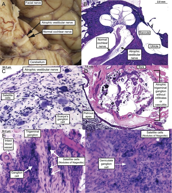
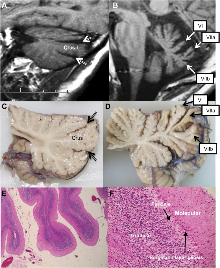
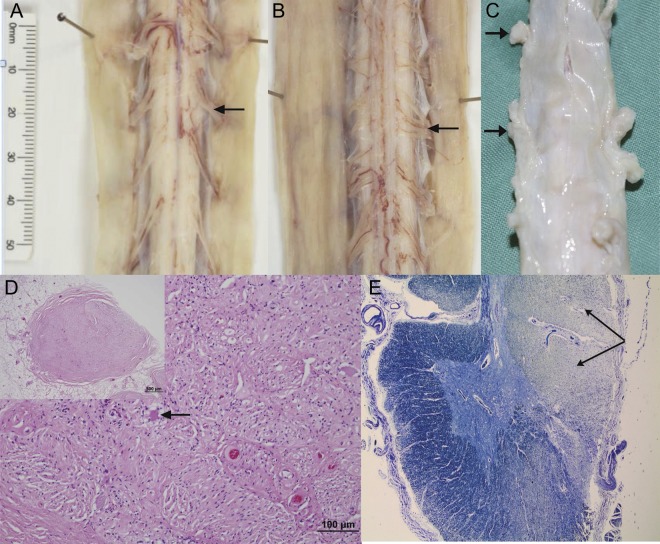
Results: Spinal cord pathology demonstrated a marked dorsal root ganglionopathy with secondary tract degeneration. Cerebellar pathology showed loss of Purkinje cells, predominantly in the vermis.

Conclusion: The likely underlying sensory pathology in CANVAS is loss of neurons from the dorsal root and V, VII, and VIII cranial nerve ganglia-in other words, it is a "neuronopathy" rather than a "neuropathy." Clinically, CANVAS is a differential diagnosis for both spinocerebellar ataxia type 3 (or Machado-Joseph disease) and Friedreich ataxia. In addition, there are 6 sets of sibling pairs, implying that CANVAS is likely to be a late-onset recessive or autosomal dominant with reduced penetrance disorder, and identification of the culprit gene is currently a target of investigation.


Figures

Figure 1
Figure 1. Temporal bone histopathology (some of this pathology has been previously reported)
(A) Marked atrophy of the vestibular component of the eighth cranial nerve in the cerebellopontine angle (compared with the adjacent cochlear component of the eighth cranial nerve). (B) Severely atrophic vestibular nerve; low-power hematoxylin & eosin (H&E). (C) Atrophic vestibular nerve with marked diminution in Scarpa ganglion cells; high-power H&E. (D) Severely atrophic trigeminal ganglion where the bulk of the ganglion has been replaced by arachnoid tissue, psammoma bodies, and CSF; low-power H&E. (E) Severely atrophic trigeminal ganglion with a marked decrease in ganglion cells, atrophy of nerve fibers, and presence of nodules of Nageotte; high-power H&E. (F) Atrophic facial nerve with near-total loss of ganglion cells; high-power H&E.
Figure 2
Figure 2. Cerebellum
(A) MRI of patient 2 showing a parasagittal pattern of atrophy predominantly affecting hemispheric crus I. Double-lined arrows indicate widening of the superior posterior and horizontal fissures. (B) MRI of patient 2 showing midsagittal pattern of atrophy predominantly affecting vermal lobules VI, VIIa, and VIIb. (C) Parasagittal cerebellum showing minor atrophy of the folia. Double-lined arrows indicate the superior posterior and horizontal fissures. (D) Midline cerebellar vermis showing marked atrophy of folia, particularly in the superior and dorsal aspect. (E) Cerebellar atrophy ×100; hematoxylin & eosin (H&E). (F) Purkinje cell loss and Bergmann layer gliosis; ×400 H&E.
Figure 3
Figure 3. Spinal cord dorsal root ganglia
(A) Normal anterior spinal cord nerve rootlets (arrow). (B) Atrophic posterior spinal cord nerve rootlets (arrow). (C) Spinal cord and coverings showing atrophic dorsal root ganglia (arrows). (D) Atrophic dorsal root ganglion showing marked neuronal loss with scant residual neurons (arrow in higher magnification main image; low-power whole dorsal root ganglion shown in inset). No evidence of satellite cell hypertrophy. (E) Cross-section of cervical spinal cord showing secondary demyelination of the posterior columns (arrows); ×20, Luxol fast blue.

References

    1. Szmulewicz DJ, Waterston JA, MacDougall HG, et al. Cerebellar ataxia, neuropathy, vestibular areflexia syndrome (CANVAS): a review of the clinical features and video-oculographic diagnosis. Ann NY Acad Sci 2011;1233:139–147 - PubMed
    1. Szmulewicz DJ, Waterston JA, Halmagyi GM, et al. Sensory neuropathy as part of the cerebellar ataxia neuropathy vestibular areflexia syndrome. Neurology 2011;76:1903–1910 - PMC - PubMed
    1. Szmulewicz DJ, Merchant SN, Halmagyi GM. Cerebellar ataxia with neuropathy and bilateral vestibular areflexia syndrome: a histopathologic case report. Otol Neurotol 2011;32:e63–e65 - PMC - PubMed
    1. Migliaccio A, Halmagyi G, Mcgarvie L, Cremer P. Cerebellar ataxia with bilateral vestibulopathy: description of a syndrome and its characteristic clinical sign. Brain 2004;127:280–293 - PubMed
    1. Koeppen AH, Morral JA, Davis AN, et al. The dorsal root ganglion in Friedreich's ataxia. Acta Neuropathol 2009;118:763–776 - PubMed

Publication types

MeSH terms

LinkOut - more resources