. 2014 May 1;192(9):4409-16.
doi: 10.4049/jimmunol.1303135. Epub 2014 Mar 28.

Cell depletion in mice that express diphtheria toxin receptor under the control of SiglecH encompasses more than plasmacytoid dendritic cells

Affiliations

Cell depletion in mice that express diphtheria toxin receptor under the control of SiglecH encompasses more than plasmacytoid dendritic cells

Melissa Swiecki et al. J Immunol. .

Abstract

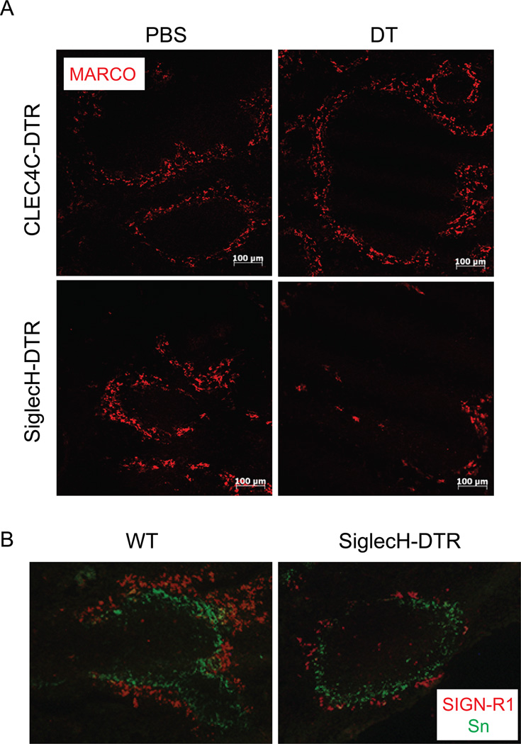
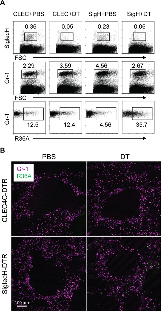
Plasmacytoid dendritic cells (pDC) produce IFN-I in response to viruses and are routinely identified in mice by SiglecH expression. SiglecH is a sialic acid-binding Ig-like lectin that has an immunomodulatory role during viral infections. In this study, we evaluated the impact of SiglecH deficiency on cytokine responses in the presence and absence of pDC. We found that lack of SiglecH enhanced IFN-I responses to viral infection, regardless of whether pDC were depleted. We also examined the expression pattern of SiglecH and observed that it was expressed by specialized macrophages and progenitors of classical dendritic cells and pDC. Accordingly, marginal zone macrophages and pDC precursors were eliminated in newly generated SiglecH-diphtheria toxin receptor (DTR)-transgenic (Tg) mice but not in CLEC4C-DTR-Tg mice after diphtheria toxin (DT) treatment. Using two bacterial models, we found that SiglecH-DTR-Tg mice injected with DT had altered bacterial uptake and were more susceptible to lethal Listeria monocytogenes infection than were DT-treated CLEC4C-DTR-Tg mice. Taken together, our findings suggest that lack of SiglecH may affect cytokine responses by cell types other than pDC during viral infections, perhaps by altering viral distribution or burden, and that cell depletion in SiglecH-DTR-Tg mice encompasses more than pDC.


Figures

Figure 1
Figure 1. Analysis of WT and SiglecH-eGFP reporter mice
(A) SiglecHeGFP/+ mice were infected or not with HSV-1 and spleens were analyzed 8 and 24 h later for pDC (Ly6C+SiglecH+eGFP+). (B) Body weights and levels of blood urea nitrogen, creatinine and total protein in blood from WT and SiglecHeGFP/eGFP mice. Data are from two independent experiments. (C) Representative light microscopy of H&E stained kidney sections revealed no pathologic abnormalities of SiglecH-deficient mice. Magnification 40×.
Figure 2
Figure 2. Effect of SiglecH deficiency on pDC responses to CpGA and MCMV
(A) pDC from WT and SiglecHeGFP/eGFP mice were enriched from BM and stimulated with CpGA for 48 h. (B) pDC from WT and SiglecHeGFP/eGFP mice were sort-purified from BM and stimulated with CpGA for 24 h. (A, B) IFN-α was measured in supernatants by ELISA. (C) WT and SiglecHeGFP/eGFP mice were injected i.v. with CpGA and serum IFN-α levels were measured 6 h later. (D) pDC were sort-purified from WT and SiglecHeGFP/eGFP mice and stimulated for 24 h with MCMV. IFN-α was measured in supernatants by ELISA. (E) WT and SiglecHeGFP/eGFP mice were infected i.p. with MCMV and serum IFN-α levels were measured 48 h p.i. Statistical significance is indicated by the p value. Data are from two (A, B, D, E) or three (C) experiments.
Figure 3
Figure 3. Lack of SiglecH impacts innate immune responses independently of pDC in vivo
WT and SiglecHeGFP/eGFP mice were infected i.v. with HSV-1. Serum IFN-α (A), TNF-α (B) and IL-6 (C) were measured 6 h p.i. (D) CLEC4C-DTR Tg mice were bred to SiglecHeGFP/+ and SiglecHeGFP/eGFP mice. Mice were injected with phosphate buffered saline (PBS) or DT 24 h before infection with HSV-1. Serum IFN-α levels were measured 6 h p.i. Data are from two or three independent experiments with at least 3 mice per group in each experiment. Statistical significance is indicated by p values.
Figure 4
Figure 4. SiglecH is expressed by specialized macrophages
(A) SiglecH is expressed by pDC and MZM in spleen. Black arrowheads denote pDC in the T cell area and red arrowheads indicate MZM. Magnification: top panels, 100× scale bar 200 micron; bottom panels, 400× scale bar 50 micron. (B) Microglia were isolated from WT mice and stained with CD45, CD11b and SiglecH. The dotplot shows CD45intCD11b+ microglia and histograms show SiglecH expression. Data are representative of two independent experiments with 3–5 mice per experiment.
Figure 5
Figure 5. SiglecH expression in progenitors of cDC and pDC
(A) CD11chi cells in SiglecHeGFP/+ mice express eGFP. The dotplot shows live spleen cells stained with CD4 and CD11c. Histograms show eGFP expression in CD4+CD11chi DC and CD4CD11chi DC in WT and SiglecHeGFP/+ mice. (B) pDC, CD8α+ and CD8α DC were sorted from spleens and SiglecH expression was measured by qPCR. (C) Precursors of pDC (pre-pDC) in BM express SiglecH. The dotplot shows B220hiSiglecH+ pDC and B220loSiglecH+ pre-pDC and histograms denote expression of CCR9, Ly6C, CD11c and MHCII on each subset. (D) Sorted pre-pDC from BM differentiate into cDC or pDC after culture in GM-CSF or Flt3L, respectively. (E) E2-2 expression in pDC, CD8α+ DC and CD8α DC sorted from spleen and mature pDC and pre-pDC sorted from BM. (F) SiglecH-DTR Tg mice, CLEC4C-DTR Tg mice and their littermates (DTR) were injected with DT and BM was analyzed for mature pDC and pre-pDC 48 h later. (A, C, F) Dotplots are representative of 5–10 mice. (B, D, E) Cells were sorted from 5–6 mice for qPCR or in vitro differentiation.
Figure 6
Figure 6. MZM are depleted in SiglecH-DTR Tg mice
(A) Spleen sections from PBS or DT-treated CLEC4C-DTR Tg mice and C57BL/6 SiglecH-DTR Tg chimeras were stained for MARCO to identify MZM (10× magnification). (B) Spleen sections from BALB/c SiglecH-DTR Tg mice and their non-Tg littermates (WT) injected with DT were stained with antibodies to SIGN-R1 and Sialoadhesin (Sn) to detect MZM and MM, respectively (20× magnification). Data are representative of two-three independent experiments.
Figure 7
Figure 7. Distribution and clearance of bacteria is altered in depleted SiglecH-DTR Tg mice
PBS or DT-treated CLEC4C-DTR Tg mice and C57BL/6 SiglecH-DTR Tg chimeras were injected with Alexa Fluor 647 labeled S. pneumoniae R36A. One hour after injection, spleens were harvested for flow cytometry and tissue sections. (A) Top panels show pDC frequencies in PBS and DT-treated mice. Middle panels show frequencies of live, Gr-1+ cells and bottom panels show frequencies of Alexa Fluor 647+ cells among live, Gr-1+ cells. (B) Spleen sections were stained with Gr-1 and show increased numbers of bacteria in the red pulp of DT-treated SiglecH-DTR Tg chimeras (10× magnification). Data are representative of three independent experiments.
Figure 8
Figure 8. Susceptibility of CLEC4C-DTR Tg mice and SiglecH-DTR Tg mice to LM-OVA
Groups of five mice: PBS or DT-treated CLEC4C-DTR Tg mice and SiglecH-DTR Tg chimeras were infected i.p. with LM-OVA. (A) Survival of mice was monitored every 12 h and (B) serum was collected from three mice in each group 24 h p.i. for cytokine analysis. Statistical significance is indicated by p values. NS, not significant.

References

    1. Gilliet M, Cao W, Liu YJ. Plasmacytoid dendritic cells: sensing nucleic acids in viral infection and autoimmune diseases. Nat Rev Immunol. 2008;8:594–606. - PubMed
    1. Trinchieri G. Type I interferon: friend or foe? J Exp Med. 2010;207:2053–2063. - PMC - PubMed
    1. Swiecki M, Gilfillan S, Vermi W, Wang Y, Colonna M. Plasmacytoid dendritic cell ablation impacts early interferon responses and antiviral NK and CD8(+) T cell accrual. Immunity. 2010;33:955–966. - PMC - PubMed
    1. Wang Y, Swiecki M, Cella M, Alber G, Schreiber RD, Gilfillan S, Colonna M. Timing and magnitude of type I interferon responses by distinct sensors impact CD8 T cell exhaustion and chronic viral infection. Cell Host Microbe. 2012;11:631–642. - PMC - PubMed
    1. Swiecki M, Wang Y, Gilfillan S, Colonna M. Plasmacytoid Dendritic Cells Contribute to Systemic but Not Local Antiviral Responses to HSV Infections. PLoS Pathog. 2013;9:e1003728. - PMC - PubMed

Publication types

MeSH terms

LinkOut - more resources