. 2013:23:62-73.
doi: 10.1007/978-3-642-38868-2_6.

Automated segmentation of the cerebellar lobules using boundary specific classification and evolution

Automated segmentation of the cerebellar lobules using boundary specific classification and evolution

John A Bogovic et al. Inf Process Med Imaging. 2013.

Abstract

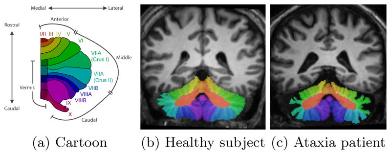
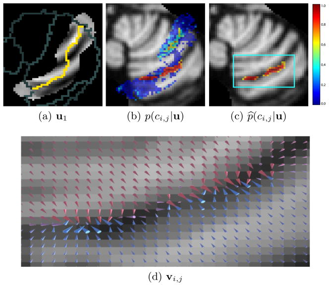
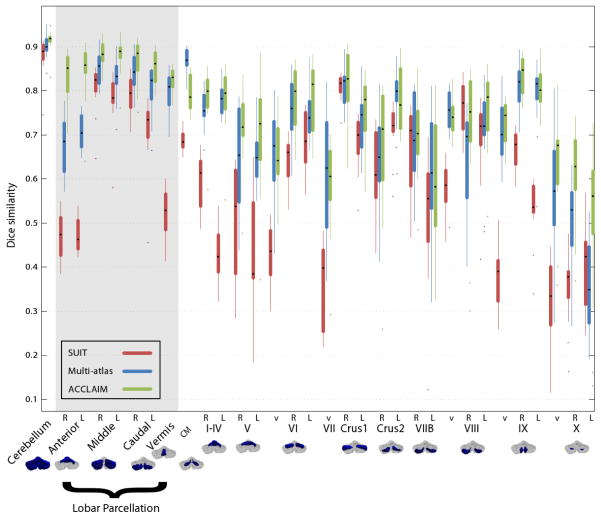
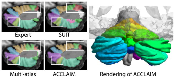
The cerebellum is instrumental in coordinating many vital functions ranging from speech and balance to eye movement. The effect of cerebellar pathology on these functions is frequently examined using volumetric studies that depend on consistent and accurate delineation, however, no existing automated methods adequately delineate the cerebellar lobules. In this work, we describe a method we call the Automatic Classification of Cerebellar Lobules Algorithm using Implicit Multi-boundary evolution (ACCLAIM). A multiple object geometric deformable model (MGDM) enables each boundary surface of each individual lobule to be evolved under different level set speeds. An important innovation described in this work is that the speed for each lobule boundary is derived from a classifier trained specifically to identify that boundary. We compared our method to segmentations obtained using the atlas-based and multi-atlas fusion techniques, and demonstrate ACCLAIM's superior performance.


Figures

Fig. 1
Fig. 1
Figures showing (a) a cartoon of the cerebellar vermis and lobules (one hemisphere), and (b) and (c) MR images of the cerebellum labeled by an expert human rater.
Fig. 2
Fig. 2
A trained classifier is given image features, one of which is shown in (a). The yellow pixels show the current estimate of the boundary to be evolved, while gray pixels show other nearby boundaries. The classifier produces a probability map shown in (b) indicating how likely it is that a given voxel lies on the boundary between two lobules. The topology of this probability map is corrected, the result of which is given in (c). Finally a GVF field is computed from the topologically correct probabilities as shown in (d). The arrows of the GVF are scaled by the square root of their magnitude and colored according to their y component: blues and reds indicate positive and negative y components, respectively.
Fig. 3
Fig. 3
Box plots Dice similarity coefficients.
Fig. 4
Fig. 4
Examples of cerebellar lobule segmenations using the SUIT, multi-atlas, and ACCLAIM methods. Shown also is a rendering of a cerebellar lobule segmentation produced by ACCLAIM, (with a transparent cerebrum for reference).

References

    1. Avants BB, Epstein CL, Grossman M, Gee JC. Symmetric diffeomorphic image registration with cross- correlation: Evaluating automated labeling of elderly and neurodegenerative brain. MIA. 2008;12(1):26–41. - PMC - PubMed
    1. Bazin PL, Ellingsen LM, Pham DL. Digital homeomorphisms in deformable registration. Proc IPMI. 2007 Jan;20:211–22. - PubMed
    1. Bazin PL, Pham DL. Topology Correction of Segmented Medical Images using a Fast Marching Algorithm. CMPB. 2007;88(2):182–190. - PMC - PubMed
    1. Bazin PL, Pham DL. Topology-preserving tissue classification of magnetic resonance brain images. IEEE TMI. 2007 May;26(4):487–96. - PubMed
    1. Bogovic JA, Prince JL, Bazin PL. A Multiple Object Geometric Deformable Model for Image Segmentation. CVIU. 2012 in press. - PMC - PubMed

Publication types

LinkOut - more resources