. 2013:23:511-23.
doi: 10.1007/978-3-642-38868-2_43.

Automatic prostate MR image segmentation with sparse label propagation and domain-specific manifold regularization

Automatic prostate MR image segmentation with sparse label propagation and domain-specific manifold regularization

Shu Liao et al. Inf Process Med Imaging. 2013.

Abstract

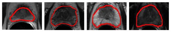
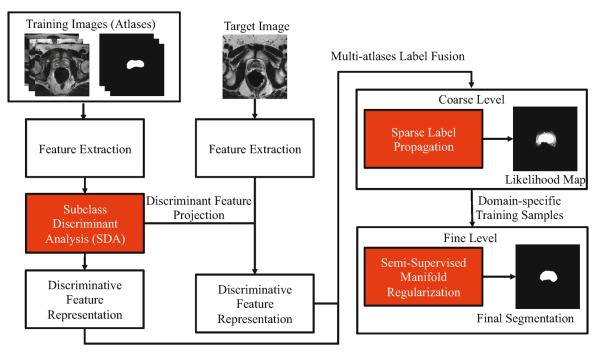
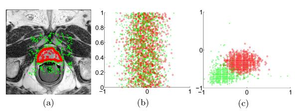
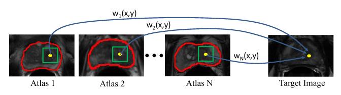
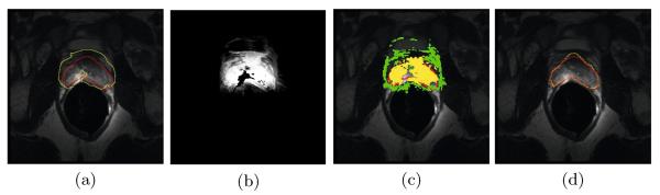
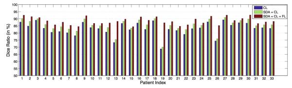
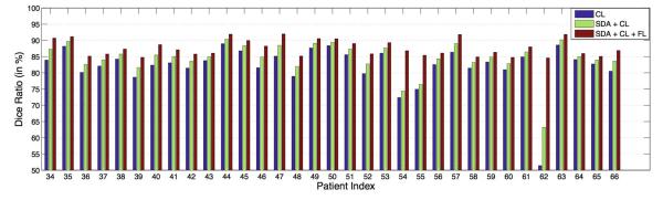
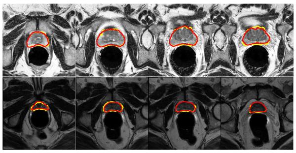
Automatic prostate segmentation in MR images plays an important role in prostate cancer diagnosis. However, there are two main challenges: (1) Large inter-subject prostate shape variations; (2) Inhomogeneous prostate appearance. To address these challenges, we propose a new hierarchical prostate MR segmentation method, with the main contributions lying in the following aspects: First, the most salient features are learnt from atlases based on a subclass discriminant analysis (SDA) method, which aims to find a discriminant feature subspace by simultaneously maximizing the inter-class distance and minimizing the intra-class variations. The projected features, instead of only voxel-wise intensity, will be served as anatomical signature of each voxel. Second, based on the projected features, a new multi-atlases sparse label fusion framework is proposed to estimate the prostate likelihood of each voxel in the target image from the coarse level. Third, a domain-specific semi-supervised manifold regularization method is proposed to incorporate the most reliable patient-specific information identified by the prostate likelihood map to refine the segmentation result from the fine level. Our method is evaluated on a T2 weighted prostate MR image dataset consisting of 66 patients and compared with two state-of-the-art segmentation methods. Experimental results show that our method consistently achieves the highest segmentation accuracies than other methods under comparison.


Figures

Fig. 1
Fig. 1
Illustrations of large inter-subject shape and appearance variations of prostate in MR images, where the manual segmentation groundtruths are highlighted by the red contours. Note also the large inhomogeneous image appearances within the prostate region.
Fig. 2
Fig. 2
Flow chart of our method, where all rectangles highlighted in red denote the main contributions of the proposed method
Fig. 3
Fig. 3
(a) A typical example of sampling voxels drawn from a training image. Samples belonging to the prostate and non-prostate regions are highlighted by small red circles and green crosses, respectively. (b) shows the scatter plot of 1D discriminant feature learnt by LDA, where the horizontal axis represents the projected value of the features. For easy visualization, the projected data points are uniformly distributed along the vertical axis. (c) shows the scatter plot of the top two most discriminant feature learnt by the SDA algorithm.
Fig. 4
Fig. 4
The schematic illustration of multi-atlases label propagation. In this example, N atlases with their segmentation groundtruths highlighted by red contours are available. For the reference voxel highlighted by the yellow dot in the target image, its prostate likelihood can be estimated by comparing its feature signature with those of the neighboring candidate voxels within the green squares in the atlases. The contribution of each candidate voxel y in the ith atlas during label propagation is determined by the graph weight wi(x, y).
Fig. 5
Fig. 5
Demonstration on how our method works on a challenging example. (a) shows the estimated prostate boundary (yellow contour) by using sparse label propagation in the coarse level, with the groundtruth prostate boundary overlayed (red contour), and (b) is the corresponding prostate likelihood map. Voxels highlighted with yellow in (c) are determined as domain-specific prostate samples, and voxels highlighted with green in (c) are determined as domain-specific non-prostate samples. (d) shows the estimated prostate boundary (yellow contour) by applying the fine level domain-specific manifold regularization, with the groundtruth prostate boundary overlayed (red contour).
Fig. 6
Fig. 6
Dice ratio between the estimated prostate volume and the groundtruth of the first 33 patients by using coarse level (CL) segmentation only, SDA derived feature with CL, and finally integrated with the fine level (FL) segmentation.
Fig. 7
Fig. 7
Dice ratio between the estimated prostate volume and the groundtruth of the rest of the 33 patients by using coarse level (CL) segmentation only, SDA derived feature with CL, and finally integrated with the fine level (FL) segmentation
Fig. 8
Fig. 8
Exemplar segmentation results obtained by the proposed method, where each row represents the segmentation results of a particular patient. Here, the estimated prostate boundary is highlighted in yellow, and the groundtruth prostate boundary is highlighted in red.

References

    1. Chandra S, Dowling J, Shen K, Raniga P, Pluim J, Greer P, Salvado O, Fripp J. Patient specific prostate segmentation in 3D magnetic resonance images. TMI. 2012;31:1955–1964. - PubMed
    1. Klein S, Heide U, Lips I, Vulpen M, Staring M, Pluim J. Automatic segmentation of the prostate in 3D MR images by atlas matching using localized mutual information. Medical Physics. 2008;35:1407–1417. - PubMed
    1. Toth R, Madabhushi A. Multi-feature landmark-free active appearance models: Application to prostate MRI segmentation. TMI. 2012;31:1638–1650. - PubMed
    1. Martin S, Troccaz J, Daanen V. Automated segmentation of the prostate in 3D MR images using a probabilistic atlas and a spatially constrained deformable model. Medical Physics. 2010;37:1579–1590. - PubMed
    1. Warfield S, Zou K, Wells W. Simultaneous truth and performance level estimation (STAPLE): An algorithm for the validation of image segmentation. TMI. 2004;23:903–921. - PMC - PubMed