Review
. 2014 Apr;171(8):2000-16.
doi: 10.1111/bph.12416.

Parthanatos: mitochondrial-linked mechanisms and therapeutic opportunities

Affiliations
Review

Parthanatos: mitochondrial-linked mechanisms and therapeutic opportunities

Amos A Fatokun et al. Br J Pharmacol. 2014 Apr.

Abstract

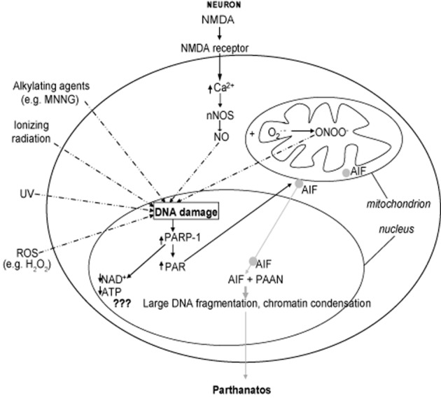
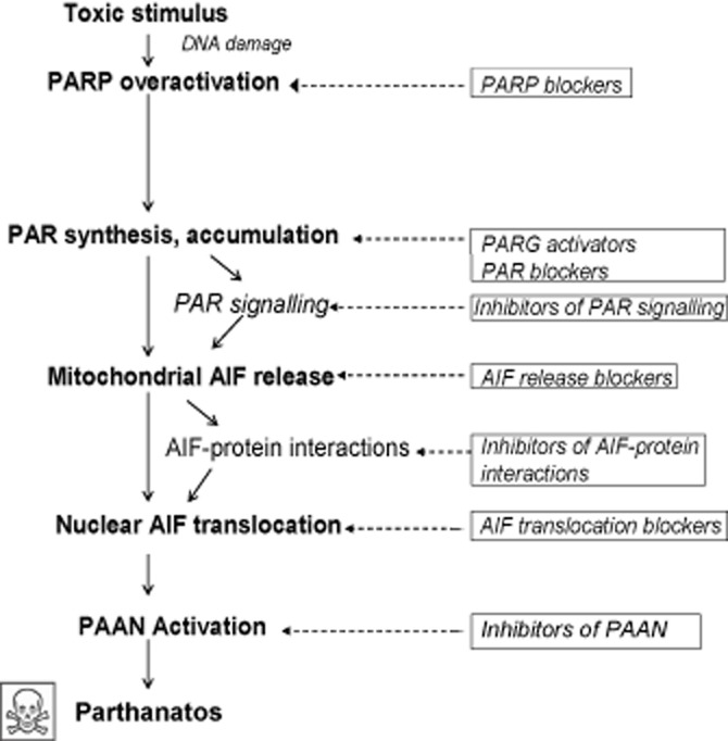
Cells die by a variety of mechanisms. Terminally differentiated cells such as neurones die in a variety of disorders, in part, via parthanatos, a process dependent on the activity of poly (ADP-ribose)-polymerase (PARP). Parthanatos does not require the mediation of caspases for its execution, but is clearly mechanistically dependent on the nuclear translocation of the mitochondrial-associated apoptosis-inducing factor (AIF). The nuclear translocation of this otherwise beneficial mitochondrial protein, occasioned by poly (ADP-ribose) (PAR) produced through PARP overactivation, causes large-scale DNA fragmentation and chromatin condensation, leading to cell death. This review describes the multistep course of parthanatos and its dependence on PAR signalling and nuclear AIF translocation. The review also discusses potential targets in the parthanatos cascade as promising avenues for the development of novel, disease-modifying, therapeutic agents.

Keywords: AIF; PARP-1; cell death; mitochondria; parthanatos; therapy.


Figures

Figure 1
Figure 1
Primary structure of PARP-1. Primary structure of PARP-1 showing its three domains: An N-terminal DNA-binding domain containing two zinc-finger motifs and a nuclear localization sequence (NLS), a central automodification domain [with a breast cancer-associated gene 1 (BRCA1) C-terminal (BRCT) motif containing phosphorylation sites for regulating PARP-1 activity], and a C-terminal catalytic domain containing the nicotinamide adenine dinucleotide (NAD)-binding site and the poly (ADP-ribose) polymer (PAR)-synthesizing domain.
Figure 2
Figure 2
Cascade of events leading to parthanatos. Diagrammatic representation, in a typical (neuronal or non-neuronal) cell, of the multiple steps in parthanatos. In neurones, activation of the NMDA receptor leads to increased calcium influx, resulting in the activation of calcium-dependent neuronal nitric oxide synthase (nNOS) that produces NO. NO may induce DNA damage directly, but more commonly reacts with superoxide (O2._) in the mitochondria to generate peroxynitrite (ONOO), which is a very potent inducer of DNA damage. Production of NO through activation of endothelial NOS (eNOS) or inducible NOS (iNOS) may be more important in non-neuronal cells, leading to ONOO generation (not shown). Some stimuli can induce DNA damage directly, including reactive oxygen species (ROS), for example hydrogen peroxide (H2O2), alkylating agents [e.g. N-methyl-N’-nitro-N-nitrosoguanidine (MNNG)], UV radiation and ionizing radiation. Stimuli inducing DNA damage are shown by broken arrows. DNA damage causes PARP-1 overactivation that leads to poly (ADP-ribose) (PAR) polymer synthesis and accumulation. PARP-1 overactivation depletes cellular pool of nicotinamide adenine dinucleotide (NAD+) and ATP, but this does not seem to be the primary cause of cell death (indicated by ???). PAR polymer signals to the mitochondria and directly binds to the PAR polymer-binding site on apoptosis-inducing factor (AIF), inducing its mitochondrial release and translocation to the nucleus. Once in the nucleus, it causes large-scale (≈50 kb) DNA fragmentation and chromatin condensation through as yet unidentified parthanatos AIF-associated nuclease (PAAN). This is believed to be the cause of cell death. Events after AIF release from the mitochondria are depicted in grey, solid arrows.
Figure 3
Figure 3
Potential therapeutic opportunities in the parthanatos cascade. Steps in the parthanatos cascade where therapeutic interventions are possible, from exposure of a cell to a toxic stimulus to the eventual cell death. Pathway progression is depicted using solid arrows. Proven or potential therapeutic interventions are in solid rectangular boxes, each linked with broken arrows to the step it modulates. To date, PARP blockers are the most advanced in development, with a number of them having entered clinical trials. The development of most other interventions is still at the experimental stage.

References

    1. Abdelkarim GE, Gertz K, Harms C, Katchanov J, Dirnagl U, Szabo C, et al. Protective effects of PJ34, a novel, potent inhibitor of poly(ADP-ribose) polymerase (PARP) in in vitro and in vivo models of stroke. Int J Mol Med. 2001;7:255–260. - PubMed
    1. Alexander SPH, Benson HE, Faccenda E, Pawson AJ, Sharman JL, Catterall WA, Spedding M, Peters JA, Harmar AJ CGTP Collaborators. The Concise Guide to PHARMACOLOGY 2013/14: Overview. Br J Pharmacol. 2013;170:1449–1867. - PMC - PubMed
    1. Ame JC, Apiou F, Jacobson EL, Jacobson MK. Assignment of the poly(ADP-ribose) glycohydrolase gene (PARG) to human chromosome 10q11.23 and mouse chromosome 14B by in situ hybridization. Cytogenet Cell Genet. 1999;85:269–270. - PubMed
    1. Andrabi SA, Kim NS, Yu SW, Wang H, Koh DW, Sasaki M, et al. Poly(ADP-ribose) (PAR) polymer is a death signal. Proc Natl Acad Sci U S A. 2006;103:18308–18313. - PMC - PubMed
    1. Andrabi SA, Dawson TM, Dawson VL. Mitochondrial and nuclear cross talk in cell death: parthanatos. Ann N Y Acad Sci. 2008;1147:233–241. - PMC - PubMed

Publication types

MeSH terms

Substances