. 2014 Apr 22;53(15):2436-41.
doi: 10.1021/bi401673b. Epub 2014 Apr 11.

Levuglandin forms adducts with histone h4 in a cyclooxygenase-2-dependent manner, altering its interaction with DNA

Affiliations

Levuglandin forms adducts with histone h4 in a cyclooxygenase-2-dependent manner, altering its interaction with DNA

Erica J Carrier et al. Biochemistry. .

Abstract

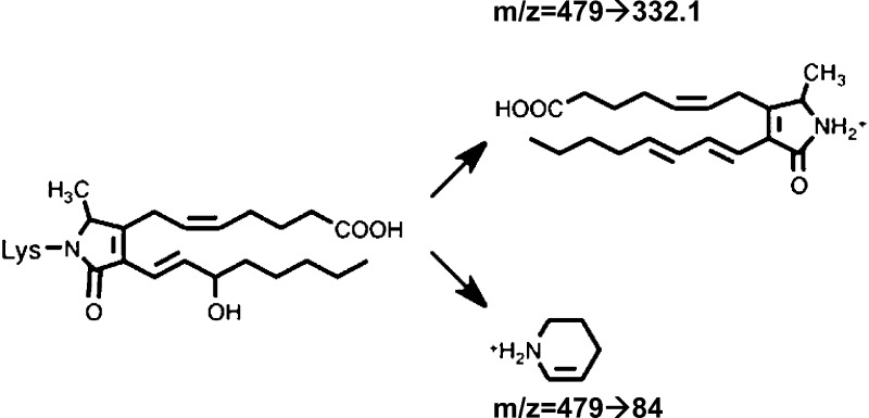
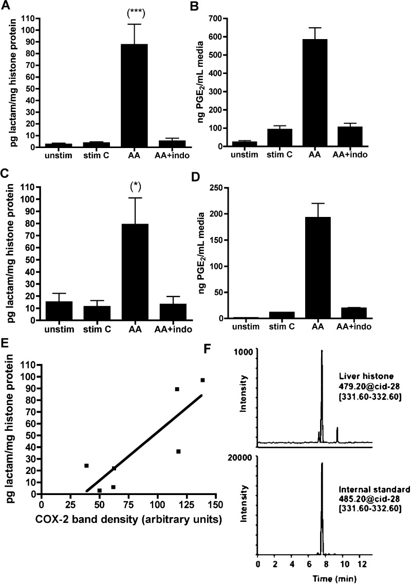
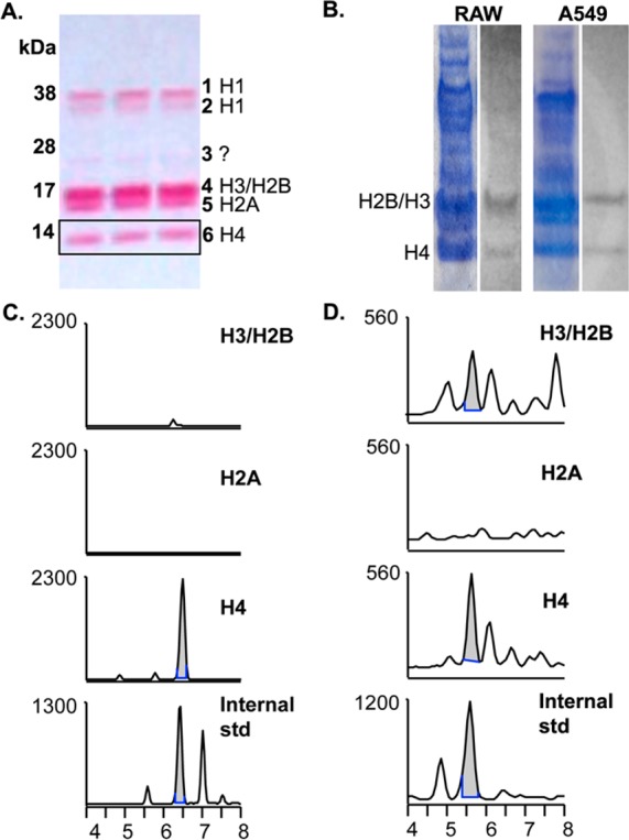
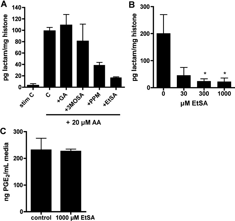
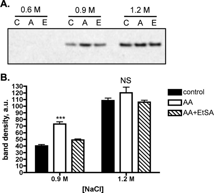
Inflammation and subsequent cyclooxygenase-2 (COX-2) activity has long been linked with the development of cancer, although little is known about any epigenetic effects of COX-2. A product of COX-2 activation, levuglandin (LG) quickly forms covalent bonds with nearby primary amines, such as those in lysine, which leads to LG-protein adducts. Here, we demonstrate that COX-2 activity causes LG-histone adducts in cultured cells and liver tissue, detectable through LC-MS, with the highest incidence in histone H4. Adduction is blocked by a γ-ketoaldehyde scavenger, which has no effect on COX-2 activity as measured by PGE2 production. Formation of the LG-histone adduct is associated with an increased histone solubility in NaCl, indicating destabilization of the nucleosome structure; this is also reversed with scavenger treatment. These data demonstrate that COX-2 activity can cause histone adduction and loosening of the nucleosome complex, which could lead to altered transcription and contribute to carcinogenesis.


Figures

Figure 1
Figure 1
Structure of the LG-lysyl adduct and the fragment ions monitored in positive ion mode (+H).
Figure 2
Figure 2
LG-lysine adducts are found in cells and tissue, dependent on COX-2 activity. RAW264.7 mouse macrophage (A) and A549 human lung carcinoma (C) cells were stimulated to express COX-2 and then given 20 μM arachidonic acid (AA) or vehicle. A subgroup of cells was preincubated 45 min with 50 μM indomethacin. As a measure of COX activity, PGE2 was determined by GC–MS from cell media prior to lysis (B and D). Nuclei were isolated, and histones were extracted and digested to individual amino acids prior to LC–ESI/MS/MS analysis. *p < 0.05; ***p < 0.001 by ANOVA followed by Tukey’s post-test (n ≥ 5). (E). Histones were extracted from nuclei of rat liver and analyzed as above for LG-lactam adduct. COX-2 protein was analyzed by Western blotting and plotted against lactam adduct levels. Each point corresponds to one liver, and shown is the line of regression (r2 = 0.7237). Pearson r = 0.8507; two-tailed p = 0.0152. (F) LC–MS chromatograph of histones isolated from a rat liver with relatively high COX-2 expression (COX-2 band intensity of 117 arbitrary units).
Figure 3
Figure 3
LG-lysyl adducts are predominantly detected on histone H4. (A). A Ponceau stain of a sample A549 histone extraction is shown, along with band identities. Histones were extracted from nuclei in 0.4 N H2SO4, resolved on 4–12% SDS-PAGE gradient gel, and transferred to nitrocellulose. H3 and H2B tend to run together as one band. (B). RAW264.7 or A549 cells were stimulated to express COX-2 and given 20 μM 14C-AA for 1 h. Cells were lysed, nuclei were isolated, and histones were extracted, concentrated, and resolved on SDS-PAGE prior to transfer to nitrocellulose and exposure to film. Shown is the Coomassie stain of the SDS-PAGE gel (left) and the result of autoradiography (right). We have observed that Ponceau, Coomassie R-250, Coomassie G-250, and silver stains each preferentially detect different histone or acid-soluble proteins. (C, D). RAW264.7 (C) or A549 (D) cells were stimulated to express COX-2 and treated with 20 μM AA for 1 h prior to histone extraction. Then 350–400 μg of total histone was loaded onto 4–12% SDS-PAGE gel and transferred to nitrocellulose. Individual bands were excised horizontally, and proteins were digested directly off the nitrocellulose by serial incubations with Pronase and aminopeptidase. The results were analyzed by LC–ESI/MS/MS, and the chromatographs of the H3/H2B and H4 bands shown against the LG-lysyl internal standard. The H2A chromatograph is shown as a representative negative result; no co-migrating peaks were seen in any other bands.
Figure 4
Figure 4
Scavenger EtSA blocks LG-lysyl adduct formation in RAW264.7 and A549 histones, without affecting COX-2 activity. (A) Scavengers were screened in RAW264.7 cells for the ability to decrease LG adduct formation on histones. Scavengers used were glucosamine (GA), 3-methoxysalicylamine (3-MoSA), pentylpyridoxamine (PPM), and 5-ethylsalicylamine (EtSA). Cells were stimulated to express COX-2, pretreated 45 min with 500 μM scavenger or vehicle (H2O), and given 20 μM AA for 1 h before lysing and extraction of histones. Histone proteins were analyzed by LC–ESI/MS/MS for LG-lysyl lactam adduct, n = 2. (B) Stimulated A549 cells were pretreated with 30, 300, or 1000 μM EtSA prior to 1 h with 20 μM AA, and histones were analyzed for LG-lysyl adduct. *p < 0.05 by one-way ANOVA followed by Dunnett’s multiple comparisons post-test (n = 3–5). (C). A549 cells were stimulated, pretreated for 45 min with 1000 μM EtSA or H2O vehicle, and given 20 μM AA for 1 h. Media was analyzed by GC–MS for PGE2 (n = 3). There was no effect on PGE2 production at lower doses of EtSA (data not shown).
Figure 5
Figure 5
LG-lysyl adduct formation on histone H4 decreases DNA–histone interaction. A549 cells were stimulated and given DMSO vehicle (C lanes) or 20 μM AA for 1 h (A lanes). A subgroup of cells was treated with 500 μM EtSA 45 min prior to adding AA (E lanes). Nuclei were extracted with 0.6, 0.9, or 1.2 M NaCl buffer, and the supernatant was evaluated by Western blotting for histone H4. Shown is a representative Western blot (A) as well as the pooled results of 4 experiments (B). Different exposure times may have been used for the 0.9 and 1.2 M bands. ***p < 0.001 by one-way ANOVA followed by Tukey’s multiple comparisons post-test. NS, not significant.

References

    1. Agrawal A.; Fentiman I. S. (2008) NSAIDs and breast cancer: a possible prevention and treatment strategy. Int. J. Clin. Pract. 62, 444–449. - PubMed
    1. Harris R. E.; Beebe-Donk J.; Alshafie G. A. (2007) Reduced risk of human lung cancer by selective cyclooxygenase 2 blockade: results of a case control study. Int. J. Biol. Sci. 3, 328–334. - PMC - PubMed
    1. Konturek P. C.; Kania J.; Burnat G.; Hahn E. G.; Konturek S. J. (2005) Prostaglandins as mediators of COX-2 derived carcinogenesis in gastrointestinal tract. J. Physiol. Pharmacol. 56(Suppl 5), 57–73. - PubMed
    1. Harris R. E. (2009) Cyclooxygenase-2 (cox-2) blockade in the chemoprevention of cancers of the colon, breast, prostate, and lung. Inflammopharmacology 17, 55–67. - PubMed
    1. Lim B. J.; Jung S. S.; Choi S. Y.; Lee C. S. (2010) Expression of metastasis-associated molecules in non-small cell lung cancer and their prognostic significance. Mol. Med. Rep. 3, 43–49. - PubMed

Publication types