

Functional characterisation of human pulmonary monocyte-like cells in lipopolysaccharide-mediated acute lung inflammation
- PMID: 24684897
- PMCID: PMC4032498
- DOI: 10.1186/1476-9255-11-9
Functional characterisation of human pulmonary monocyte-like cells in lipopolysaccharide-mediated acute lung inflammation
Abstract
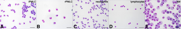
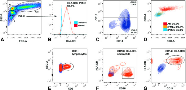
Background: We have previously reported the presence of novel subpopulations of pulmonary monocyte-like cells (PMLC) in the human lung; resident PMLC (rPMLC, HLA-DR(+)CD14(++)CD16(+)cells) and inducible PMLC (iPMLC, HLA-DR(+)CD14(++)CD16(-) cells). iPMLC are significantly increased in bronchoalveolar lavage (BAL) fluid following inhalation of lipopolysaccharide (LPS). We have carried out the first functional evaluation of PMLC subpopulations in the inflamed lung, following the isolation of these cells, and other lineages, from BAL fluid using novel and complex protocols.
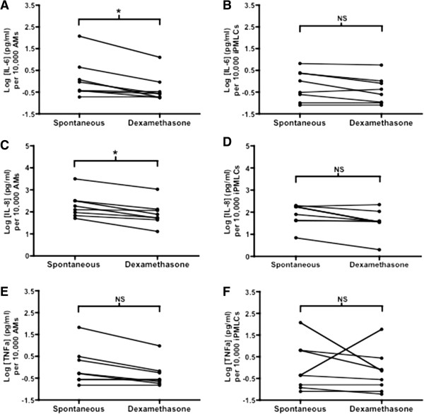
Methods: iPMLC, rPMLC, alveolar macrophages (AM), neutrophils, and regulatory T cells were quantified in BAL fluid of healthy subjects at 9 hours post-LPS inhalation (n = 15). Cell surface antigen expression by iPMLC, rPMLC and AM and the ability of each lineage to proliferate and to undergo phagocytosis were investigated using flow cytometry. Basal cytokine production by iPMLC compared to AM following their isolation from BAL fluid and the responsiveness of both cell types following in vitro treatment with the synthetic corticosteroid dexamethasone were assessed.
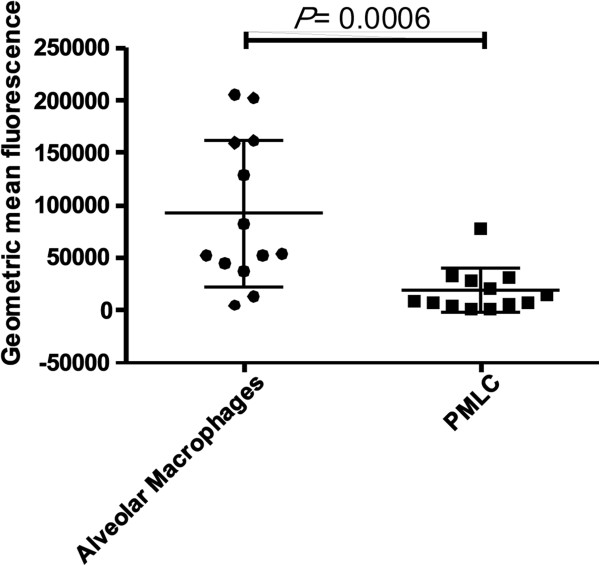
Results: rPMLC have a significantly increased expression of mature macrophage markers and of the proliferation antigen Ki67, compared to iPMLC. Our cytokine data revealed a pro-inflammatory, corticosteroid-resistant phenotype of iPMLC in this model.
Conclusions: These data emphasise the presence of functionally distinct subpopulations of the monocyte/macrophage lineage in the human lung in experimental acute lung inflammation.
Keywords: Acute lung inflammation; Corticosteroid; Lipopolysaccharide; Macrophages; Monocytes; Multiparameter flow cytometry.
Figures
References
-
- Barr LC, Brittan M, Morris AC, McAuley DF, McCormack C, Fletcher AM, Richardson H, Connell M, Patel D, Wallace WA, Rossi AG, Davidson DJ, Manson L, Turner M, Hirani N, Walsh TS, Anderson NH, Dhaliwal K, Simpson AJ. An RCT of peripheral blood mononuclear cell depletion in experimental human lung inflammation. Am J Respir Crit Care Med. 2013;188(4):449–55. doi: 10.1164/rccm.201212-2334OC. - DOI - PubMed
-
- Brittan M, Barr L, Conway Morris A, Duffin R, Rossi F, Johnston S, Monro G, Anderson N, Rossi AG, McAuley DF, Haslett C, Hirani N, Dhaliwal K, Simpson AJ. A novel subpopulation of monocyte-like cells in the human lung after lipopolysaccharide inhalation. Eur Respir J. 2012;40(1):206–214. doi: 10.1183/09031936.00113811. - DOI - PubMed
-
- Rosseau S, Hammerl P, Maus U, Walmrath HD, Schütte H, Grimminger F, Seeger W, Lohmeyer J. Phenotypic characterization of alveolar monocyte recruitment in acute respiratory distress syndrome. Am J Physiol Lung Cell Mol Physiol. 2000;279(1):L25–L35. - PubMed
-
- Hance AJ, Douches S, Winchester RJ, Ferrans VJ, Crystal RG. Characterization of mononuclear phagocyte subpopulations in the human lung by using monoclonal antibodies: changes in alveolar macrophage phenotype associated with pulmonary sarcoidosis. J Immunol. 1985;134(1):284–292. - PubMed
Grants and funding
LinkOut - more resources
Full Text Sources
Other Literature Sources
Research Materials
