. 2014 Apr 17;40(4):594-607.
doi: 10.1016/j.immuni.2014.03.005. Epub 2014 Mar 27.

Segmented filamentous bacteria antigens presented by intestinal dendritic cells drive mucosal Th17 cell differentiation

Affiliations

Segmented filamentous bacteria antigens presented by intestinal dendritic cells drive mucosal Th17 cell differentiation

Yoshiyuki Goto et al. Immunity. .

Abstract

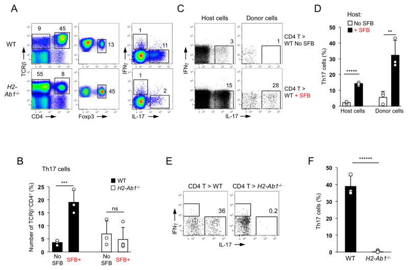
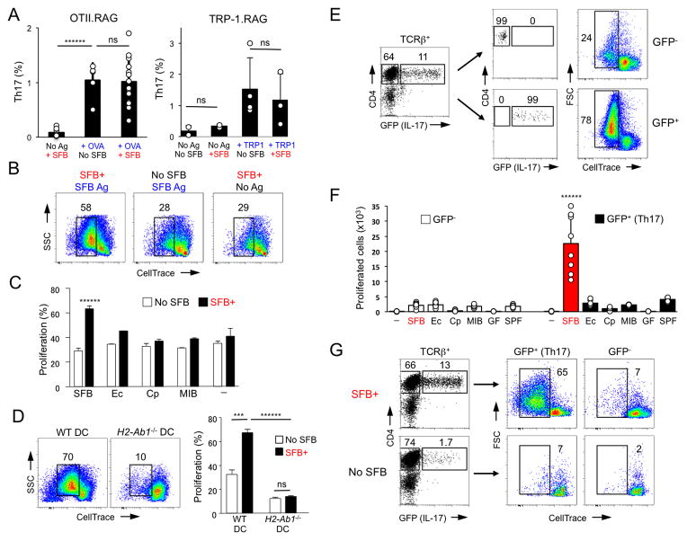
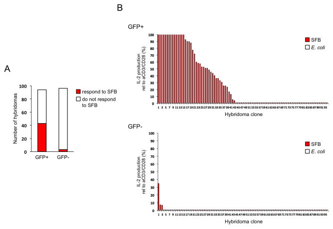
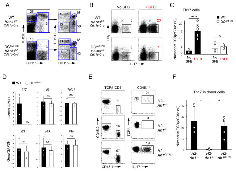
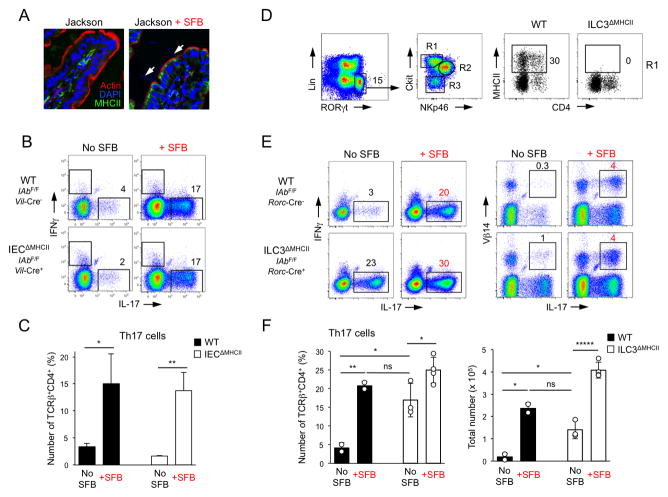
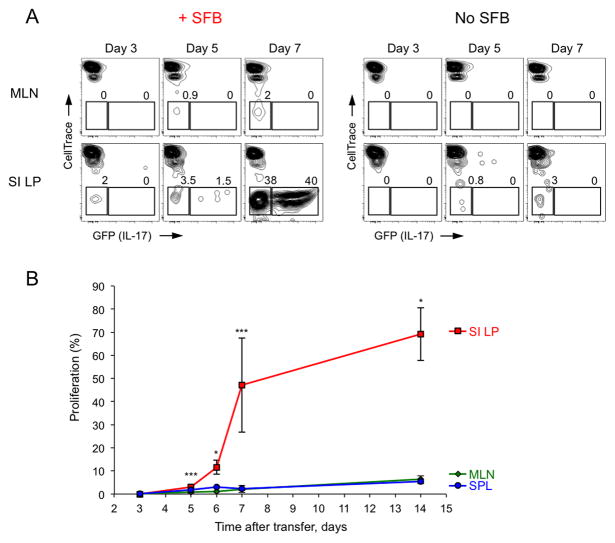
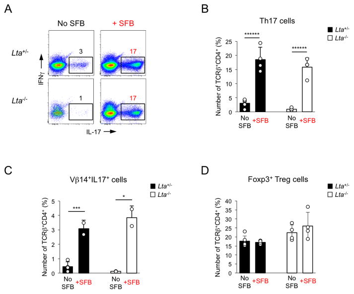
How commensal microbiota contributes to immune cell homeostasis at barrier surfaces is poorly understood. Lamina propria (LP) T helper 17 (Th17) cells participate in mucosal protection and are induced by commensal segmented filamentous bacteria (SFB). Here we show that MHCII-dependent antigen presentation of SFB antigens by intestinal dendritic cells (DCs) is crucial for Th17 cell induction. Expression of MHCII on CD11c(+) cells was necessary and sufficient for SFB-induced Th17 cell differentiation. Most SFB-induced Th17 cells recognized SFB in an MHCII-dependent manner. SFB primed and induced Th17 cells locally in the LP and Th17 cell induction occurred normally in mice lacking secondary lymphoid organs. The importance of other innate cells was unveiled by the finding that MHCII deficiency in group 3 innate lymphoid cells (ILCs) resulted in an increase in SFB-independent Th17 cell differentiation. Our results outline the complex role of DCs and ILCs in the regulation of intestinal Th17 cell homeostasis.


Figures

Figure 1
Figure 1. Induction of intestinal Th17 cells by SFB requires MHCII expression in the periphery
A. Th17 and Treg cell proportions in SI LP of SFB-positive WT and MHCII-deficient (IAb−/−) mice. Foxp3 and cytokine staining plots are gated on TCRβ+CD4+ cells B. Th17 and Treg cell proportions in SI LP of SFB-negative (Jackson microbiota) and SFB-positive (Taconic microbiota) WT and IAb−/− mice. Plots gated on TCRβ+CD4+ cells C–D. WT CD45.1+ CD4 T cells were adoptively transferred into WT CD45.2 mice before or 12 days after SFB colonization. Cytokine expression in host (CD45.2+) and donor (CD45.1+) SI LP TCRβ+CD4+ cells 2 weeks after transfer. Data from one of multiple experiments E–F. Th17 cell induction in WT CD4 T cells two weeks after transfer into SFB-positive WT and MHCII-deficient recipients. Plots gated on TCRβ+CD4+ cells. Data from one of multiple experiments
Figure 2
Figure 2. SFB-induced intestinal Th17 cells preferentially respond to SFB antigens
A. Th17 cell proportions in the SI LP of OTII.RAG and TRP-1.RAG TCR Tg mice before and after SFB colonization in the absence or presence of cognate antigen. Representative data from 5 independent experiments B–C. Proliferation response of sorted SI LP TCRβ+CD4+ cells from SFB-negative (Jax) and SFB-positive (Tac) WT B6 mice to SFB (B, C) or other bacterial antigens (C). T cell proliferation was scored by dye dilution on Day 3. Ec, E. coli, Cp, Clostridium perfringens; MIB, mouse intestinal bacteria (cultured isolates from feces of SFB-negative (Jackson) mice); “-” – no antigen. Representative data from 5 independent experiments D. SI LP TCRβ+CD4+ cells were purified from SFB-negative (No SFB) and SFB-positive (SFB+) WT mice and co-cultured with SFB antigens as in (B) and WT or IAb−/− DCs. Data from 2 independent experiments E–F. SI LP GFP+ (Th17) and GFP (non-Th17) TCRβ+CD4+ cells from SFB-positive Il17GFP mice were stimulated in vitro with SFB (E, F) or various bacterial antigens (F) as in (B) or with lysates from germ-free (GF) or SFB-negative SPF (SPF) animals. Representative data from multiple experiments G. SI LP GFP+ (Th17) and GFP (non-Th17) TCRβ+CD4+ cells from SFB-positive (SFB+) or SFB-negative (No SFB) Il17GFP mice were stimulated in vitro with SFB antigens as in (B). Representative data from 2 independent experiments
Figure 3
Figure 3. Most intestinal SFB-induced Th17 cells recognize SFB
T cell hybridomas were generated from SI LP GFP+ (Th17) and GFP (non-Th17) CD4 T cells from SFB-positive Il17GFP mice. Data combined from 2 independent experiments A. Number of hybridomas responding to SFB B. Response of individual hybridomas (percentage of maximum anti-CD3 and anti-CD28 stimulation) to SFB or E. coli antigens as assessed by IL-2 production. Clones were ordered in decreasing amounts of IL-2 production
Figure 4
Figure 4. DC expression of MHCII is necessary and sufficient for SFB-mediated Th17 cell induction
A. SI LP lymphocytes from DCΔMHCII and control littermates. Left panels, gated on TCRβCD4 cells. Right panels, gated on CD11c+ cells B–C. Th17 cell induction in DCΔMHCII mice and control littermates 2 weeks after SFB colonization. Plots gated on TCRβ+CD4+ cells. Representative data from 4 independent experiments D. Relative cytokine expression (RT-PCR) in terminal ileum of DCΔMHCII and control littermates (WT) 2 weeks after colonization with SFB. nd – below threshold of detection E–F. Th17 cell differentiation of WT CD45.1+ CD4 T cells in the SI LP 2 weeks after transfer into SFB-positive IAb+/−, IAb −/−, and IAbCD11c CD45.2+ recipient littermates. Data from 2 independent experiments
Figure 5
Figure 5. RORγt+ ILCs inhibit differentiation of SFB-independent intestinal Th17 cells through MHCII
A. MHCII expression on IECs in Jackson B6 mice before and 2 weeks after SFB colonization. Arrows point to SFB filaments attaching to IECs B–C. Th17 cell induction in IECΔMHCII mice and control littermates 2 weeks after SFB colonization. Plots gated on TCRβ+CD4+ cells. Data from one of 2 independent experiments D. Expression of MHCII on SI LP c-kit+NKp46RORγt+ group 3 ILCs in ILC3ΔMHCII mice and control littermates E–F. Th17 cell induction in ILC3ΔMHCII and control littermates 2 weeks after SFB colonization. Plots gated on TCRβ+CD4+ cells. Data from 2 independent experiments
Figure 6
Figure 6. Priming and induction of Th17 cells by SFB occurs in the small intestine
CellTrace Violet labeled CD45.2+ CD4 T cells from Il17GFP mice were transferred into WT CD45.1+ recipients before (No SFB) or after (+SFB) SFB colonization A–B. Proliferation (A, B) and Th17 cell induction (A) at indicated time points. Plots are gated on CD45.2+TCRβ+CD4+ transferred cells. SI LP, small intestinal LP; MLN, mesenteric lymph nodes; SPL, spleen. Combined data from 3 independent experiments
Figure 7
Figure 7. SFB induce Th17 cells in the absence of secondary lymphoid organs
SI LP lymphocytes were isolated from Lta−/− and control littermates 2 weeks after colonization with SFB A–C. Th17 and Vβ14+IL-17+ cells induction in TCRβ+CD4+ cells. Representative data from one of 2 independent experiments D. Foxp3+ Treg cell proportions in TCRβ+CD4+ cells. Combined data from 2 independent experiments

Comment in

References

    1. Arpaia N, Campbell C, Fan X, Dikiy S, van der Veeken J, deRoos P, Liu H, Cross JR, Pfeffer K, Coffer PJ, Rudensky AY. Metabolites produced by commensal bacteria promote peripheral regulatory T-cell generation. Nature. 2013;504:451–455. - PMC - PubMed
    1. Atarashi K, Nishimura J, Shima T, Umesaki Y, Yamamoto M, Onoue M, Yagita H, Ishii N, Evans R, Honda K, Takeda K. ATP drives lamina propria T(H)17 cell differentiation. Nature. 2008;455:808–812. - PubMed
    1. Atarashi K, Tanoue T, Oshima K, Suda W, Nagano Y, Nishikawa H, Fukuda S, Saito T, Narushima S, Hase K, et al. Treg induction by a rationally selected mixture of Clostridia strains from the human microbiota. Nature. 2013;500:232–236. - PubMed
    1. Atarashi K, Tanoue T, Shima T, Imaoka A, Kuwahara T, Momose Y, Cheng G, Yamasaki S, Saito T, Ohba Y, et al. Induction of colonic regulatory T cells by indigenous Clostridium species. Science. 2011;331:337–341. - PMC - PubMed
    1. Barnden MJ, Allison J, Heath WR, Carbone FR. Defective TCR expression in transgenic mice constructed using cDNA-based alpha- and beta-chain genes under the control of heterologous regulatory elements. Immunology and cell biology. 1998;76:34–40. - PubMed

Publication types

MeSH terms

Substances