. 2014 Jun;5(4):415-22.
doi: 10.1016/j.ttbdis.2014.01.010. Epub 2014 Mar 29.

Coinfection of tick cell lines has variable effects on replication of intracellular bacterial and viral pathogens

Affiliations

Coinfection of tick cell lines has variable effects on replication of intracellular bacterial and viral pathogens

Anna Moniuszko et al. Ticks Tick Borne Dis. 2014 Jun.

Abstract

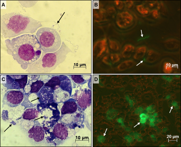
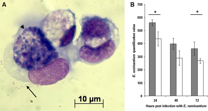
Ticks transmit various human and animal microbial pathogens and may harbour more than one pathogen simultaneously. Both viruses and bacteria can trigger, and may subsequently suppress, vertebrate host and arthropod vector anti-microbial responses. Microbial coinfection of ticks could lead to an advantage or disadvantage for one or more of the microorganisms. In this preliminary study, cell lines derived from the ticks Ixodes scapularis and Ixodes ricinus were infected sequentially with 2 arthropod-borne pathogens, Borrelia burgdorferi s.s., Ehrlichia ruminantium, or Semliki Forest virus (SFV), and the effect of coinfection on the replication of these pathogens was measured. Prior infection of tick cell cultures with the spirochaete B. burgdorferi enhanced subsequent replication of the rickettsial pathogen E. ruminantium whereas addition of spirochaetes to cells infected with E. ruminantium had no effect on growth of the latter. Both prior and subsequent presence of B. burgdorferi also had a positive effect on SFV replication. Presence of E. ruminantium or SFV had no measurable effect on B. burgdorferi growth. In tick cells infected first with E. ruminantium and then with SFV, virus replication was significantly higher across all time points measured (24, 48, 72h post infection), while presence of the virus had no detectable effect on bacterial growth. When cells were infected first with SFV and then with E. ruminantium, there was no effect on replication of either pathogen. The results of this preliminary study indicate that interplay does occur between different pathogens during infection of tick cells. Further study is needed to determine if this results from direct pathogen-pathogen interaction or from effects on host cell defences, and to determine if these observations also apply in vivo in ticks. If presence of one pathogen in the tick vector results in increased replication of another, this could have implications for disease transmission and incidence.

Keywords: Borrelia; Coinfection; Ehrlichia; Ixodes spp.; Semliki Forest virus; Tick cell line.


Figures

Fig. 1
Fig. 1
ISE6 cell cultures inoculated with B. burgdorferi s.s. (KS20), E. ruminantium (Ball 3), or Semliki Forest virus (SFV4-steGFP). (A) Giemsa-stained cytocentrifuge smear of ISE6 cells with associated B. burgdorferi (KS20) spirochaetes (arrow) at 24 h p.i.; (B) live ISE6 cells with B. burgdorferi (KS20) spirochaetes (arrows) at 6 h p.i. viewed with simultaneous brightfield and UV light; (C) Giemsa-stained cytocentrifuge smear of ISE6 cells infected with E. ruminantium (Ball3) (arrows) at 168 h p.i.; (D) live ISE6 cells infected with SFV4-steGFP at 24 h p.i. viewed with simultaneous brightfield and UV light; arrows indicate cells producing eGFP as a result of virus infection.
Fig. 2
Fig. 2
Infection of ISE6 cells with B. burgdorferi s.s. and E. ruminantium. (A) B. burgdorferi spirochaete (arrow) “interacting” with an ISE6 cell containing a colony of E. ruminantium (arrowhead). (B) Quantification of E. ruminantium in ISE6 cells infected with B. burgdorferi followed 24 h later by E. ruminantium. Shaded bars: cells infected with B. burgdorferi and E. ruminantium; white bars: cells infected with E. ruminantium alone. The amount of E. ruminantium DNA detected by real-time PCR was significantly higher in cells with B. burgdorferi than in cells with E. ruminantium alone at 24 and 72 h p.i. (Student's t-test) and across all time points (ANOVA, p = 0.0012). Experiment was repeated twice with similar results; 3 replicates per treatment, error bars represent standard deviation. * indicates p value <0.05 by Student's t-test.
Fig. 3
Fig. 3
Renilla luciferase activity in tick cells infected with a bacterium and SFV4(3H)-Rluc (Semliki Forest virus). Shaded bars: cells infected with a bacterium and SFV; white bars: cells infected with SFV alone. Experiments were repeated twice with similar results; 3 replicates per treatment, error bars represent standard deviation. (A) IRE/CTVM19 cells infected with B. burgdorferi s.s. followed 24 h later by SFV4(3H)-Rluc. Luciferase activity peaked in all cultures at 24 h p.i., and there was an increase in activity of cells with B. burgdorferi, but it was not significant (Student's t-test, p = 0.07). The difference in activity between cells with and without B. burgdorferi was not significant over all time points (ANOVA). (B) Renilla luciferase activity in IRE/CTVM19 cells infected with SFV4(3H)-Rluc followed 48 h later by B. burgdorferi. Luciferase activity was significantly higher 6 h after addition of B. burgdorferi to SFV-infected cells compared to cells with SFV alone (Student's t-test, p < 0.05), while by ANOVA there was no significant difference across all time points. (C) Renilla luciferase activity in ISE6 cells infected with E. ruminantium followed 7 days later by SFV4(3H)-Rluc. Luciferase activity was highest in all cultures at 24 h p.i. with significantly higher activity in cells coinfected with E. ruminantium and SFV compared to cells infected with SFV alone at 24 and 48 h p.i. (Student's t-test) and across all time points (ANOVA, p = 0.0028). * indicates a p value <0.05 by Student's t-test.

References

    1. Alekseev A.N., Semenov A.V., Dubinina H.V. Evidence of Babesia microti infection in multi-infected Ixodes persulcatus ticks in Russia. Exp. Appl. Acarol. 2003;29:345–353. - PubMed
    1. Alekseev A.N., Dubinina H.V., Van De Pol I., Schouls L.M. Identification of Ehrlichia spp. and Borrelia burgdorferi in Ixodes ticks in the Baltic regions of Russia. J. Clin. Microbiol. 2001;39:2237–2242. - PMC - PubMed
    1. Ayllon N., Villar M., Busby A.T., Kocan K.M., Blouin E.F., Bonzon-Kulichenko E., Galindo R.C., Mangold A.T., Alberdi P., Perez de la Lastra J.M., Vazquez J., de la Fuente J. Anaplasma phagocytophilum inhibits apoptosis and promotes cytoskeleton rearrangement for infection of tick cells. Infect. Immun. 2013;81:2415–2425. - PMC - PubMed
    1. Babb K., McAlister J.D., Miller J.C., Stevenson B. Molecular characterization of Borrelia burgdorferi erp promoter/operator elements. J. Bacteriol. 2004;186:2745–2756. - PMC - PubMed
    1. Barry G., Alberdi P., Schnettler E., Weisheit S., Kohl A., Fazakerley J.K., Bell-Sakyi L. Gene silencing in tick cell lines using small interfering or long double-stranded RNA. Exp. Appl. Acarol. 2013;59:319–338. - PMC - PubMed

Publication types