

Cognitive, genetic, and brain perfusion factors associated with four year incidence of Alzheimer's disease from mild cognitive impairment
- PMID: 24685632
- PMCID: PMC4111792
- DOI: 10.3233/JAD-132516
Cognitive, genetic, and brain perfusion factors associated with four year incidence of Alzheimer's disease from mild cognitive impairment
Abstract
Background: There is a range of factors that predict the development of Alzheimer's disease (AD) dementia among patients with amnestic mild cognitive impairment (MCI).
Objectives: To identify the neuropsychological, genetic, and functional brain imaging data that best predict conversion to AD dementia in patients with amnestic MCI.
Methods: From an initial group of 42 amnestic MCI patients assessed with neurological, neuropsychological, and brain SPECT, 39 (25 converters, 14 non-converters) were followed for 4 years, and 36 had APOE ε4 genotyping. Baseline neuropsychological data and brain SPECT data were used to predict which of the MCI patients would develop dementia by the end of the 4 years of observation.
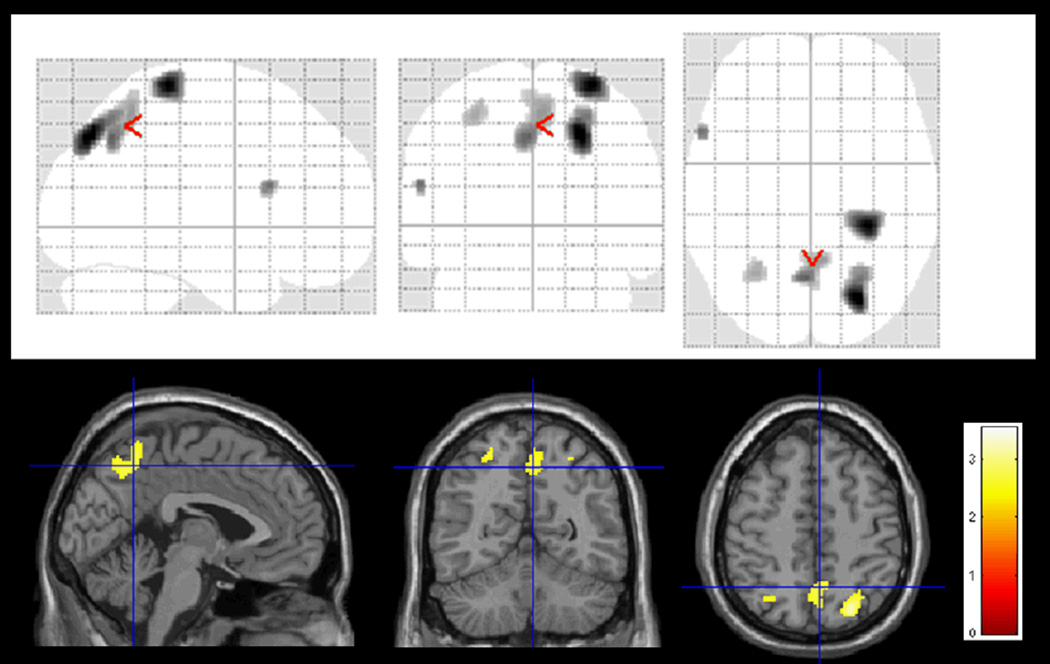
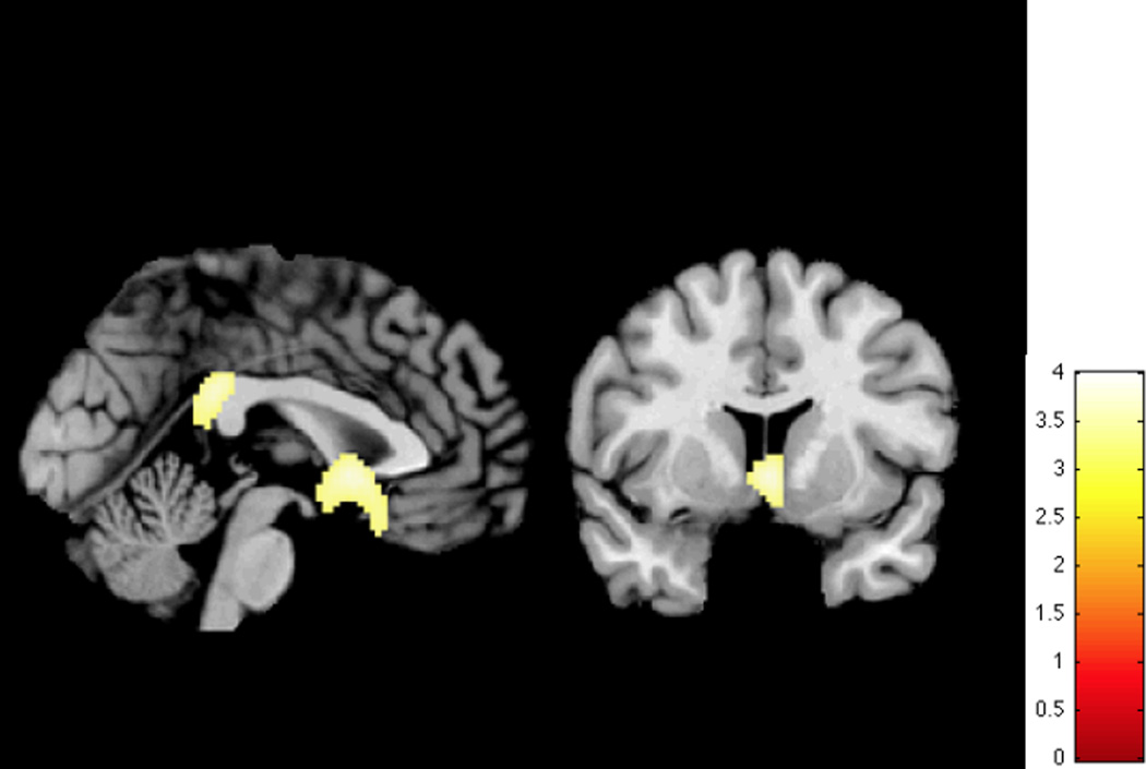
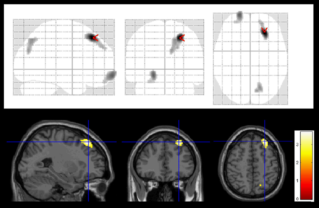
Results: The MCI patients who had converted to AD dementia had poorer performance on long-term visual memory and Semantic Fluency tests. The MCI subjects who developed dementia were more likely to carry at least one copy of the APOE ε4 allele (Hazard Risk = 4.22). There was lower brain perfusion in converters than non-converters, mainly in postcentral gyrus. An additional analysis of the SPECT data found differences between the MCI subjects and controls in the posterior cingulate gyrus and the basal forebrain. When the brain imaging and neuropsychological test data were combined in the same Cox regression model, only the neuropsychological test data were significantly associated with time to dementia.
Conclusion: Although the presence of reduced brain perfusion in postcentral gyrus and basal forebrain indicated an at-risk condition, it was the extent of memory impairment that was linked to the speed of decline from MCI to AD.
Keywords: Alzheimer's disease; brain SPECT; cerebral perfusion; four-year follow-up; longitudinal; mild cognitive impairment; prospective; visual memory.
Figures
References
-
- Morris JC. Early-stage and preclinical Alzheimer disease. Alzheimer Dis Assoc Disord. 2005;19:163–165. - PubMed
-
- Jack CR, Jr, Knopman DS, Jagust WJ, Petersen RC, Weiner MW, Aisen PS, Shaw LM, Vemuri P, Wiste HJ, Weigand SD, Lesnick TG, Pankratz VS, Donohue MC, Trojanowski JQ. Tracking pathophysiological processes in Alzheimer's disease: an updated hypothetical model of dynamic biomarkers. Lancet Neurol. 2013;12:207–216. - PMC - PubMed
-
- Sperling RA, Aisen PS, Beckett LA, Bennett DA, Craft S, Fagan AM, Iwatsubo T, Jack CR, Jr, Kaye J, Montine TJ, Park DC, Reiman EM, Rowe CC, Siemers E, Stern Y, Yaffe K Carrillo MC, Thies B, Morrison-Bogarad M, Wgster MV, Phelps CH. Toward defining the preclinical stages of Alzheimer’s disease: Recommendations from the National Institute on Aging-Alzheimer’s Association workgroups on diagnostic guidelines for Alzheimer’s disease. Alzheimers Dement. 2011;7:280–292. - PMC - PubMed
Publication types
MeSH terms
Substances
Grants and funding
LinkOut - more resources
Full Text Sources
Other Literature Sources
Medical
Miscellaneous
