. 2014 Oct;10(7):1517-27.
doi: 10.1016/j.nano.2014.03.014. Epub 2014 Mar 29.

Exosome-mediated delivery of functionally active miRNA-155 inhibitor to macrophages

Affiliations

Exosome-mediated delivery of functionally active miRNA-155 inhibitor to macrophages

Fatemeh Momen-Heravi et al. Nanomedicine. 2014 Oct.

Abstract

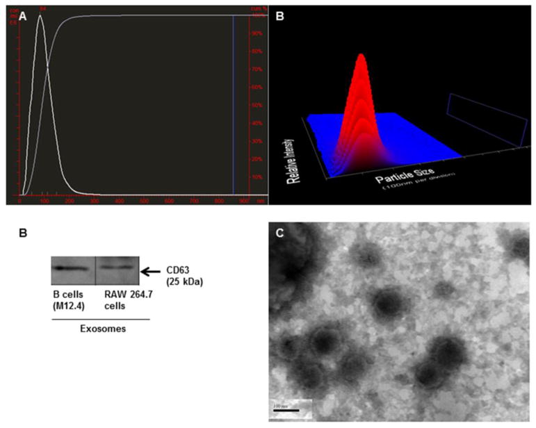
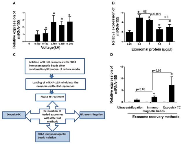
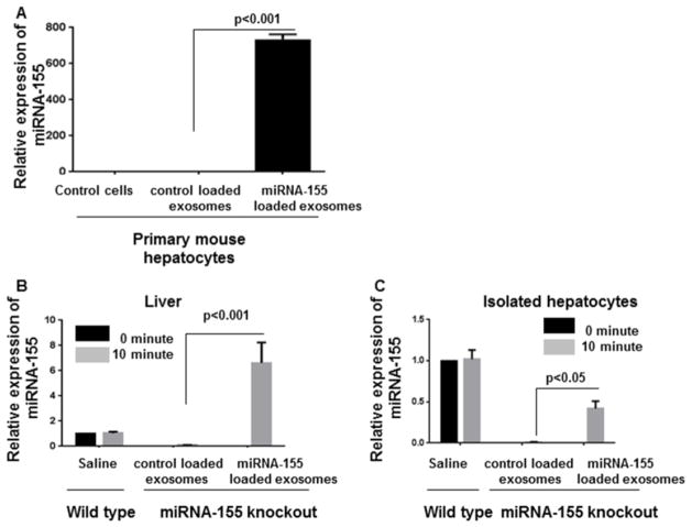
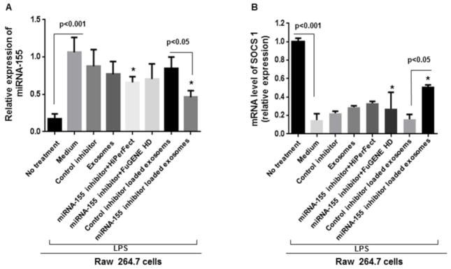
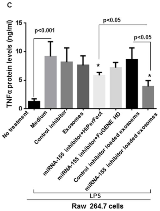
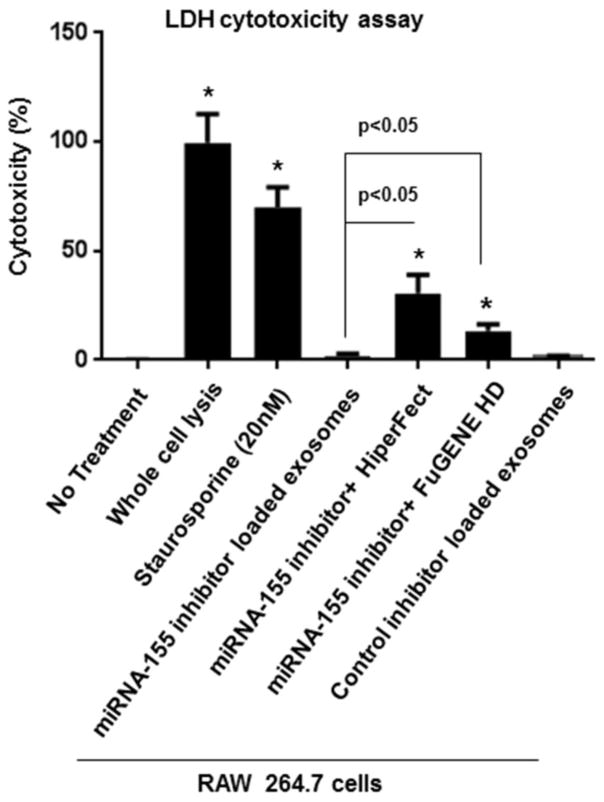
Exosomes, membranous nanovesicles, naturally carry bio-macromolecules and play pivotal roles in both physiological intercellular crosstalk and disease pathogenesis. Here, we showed that B cell-derived exosomes can function as vehicles to deliver exogenous miRNA-155 mimic or inhibitor into hepatocytes or macrophages, respectively. Stimulation of B cells significantly increased exosome production. Unlike in parental cells, baseline level of miRNA-155 was very low in exosomes derived from stimulated B cells. Exosomes loaded with a miRNA-155 mimic significantly increased miRNA-155 levels in primary mouse hepatocytes and the liver of miRNA-155 knockout mice. Treatment of RAW macrophages with miRNA-155 inhibitor loaded exosomes resulted in statistically significant reduction in LPS-induced TNFα production and partially prevented LPS-induced decrease in SOCS1 mRNA levels. Furthermore, exosome-mediated miRNA-155 inhibitor delivery resulted in functionally more efficient inhibition and less cellular toxicity compared to conventional transfection methods. Similar approaches could be useful in modification of target biomolecules in vitro and in vivo. From the clinical editor: In this study, exosome-based delivery of miRNA-155 mimicker or inhibitor was found to have significant biological response in hepatocytes and macrophages. Exosome-based approaches may be useful in the modification of other target biomolecules.

Keywords: Exosomes; Macrophages; RNAs delivery; TNFα; miRNA-155.


Conflict of interest statement

Conflict of interest: The authors have declared that no conflict of interest exists.

Figures

Figure 1
Figure 1. Charachterization of exosomes
(A) Average size of stimulated B cell-derived exosomes was 98nm with the mode of 84nm. 3D graph represents particle size versus intensity versus concentration (particles/ml) of stimulated B cell-derived exosomes. (B) Isolated exosomes from murine B cells (M12.4) and RAW 264.7 cells expressed exosomal marker, CD63. (C) TEM image of B cell exosomes showed the diameter of less than 150 nm.
Figure 2
Figure 2. Stimulation of B cell exosomes and characterization of miRNA-155 profile
(A) The exosome production was increased in B cells upon stimulation with IL-4 and CD40. The number of exosomes was quantified using NanoSight. (B) Baseline levels of miRNA-155 in different groups including RAW macrophages, non-stimulated B cells, stimulated B cells with IL-4 &CD 40, and pertinent exosomes are shown. Although a considerable amount of miRNA-155 was present in the parental B cells, they were not sorted into the exosomes (p<0.05). Exosomes derived from stimulated B cells showed statistically significant lower levels of miRNA-155 compared to other experimental groups (p<0.001). The results represent three independent experiments. (*indicates p < 0.05 versus parental cells)
Figure 3
Figure 3. Optimization of loading of miRNA-155 mimic to the exosomes
(A) miRNA-155 mimic was introduced to the exosomes using various voltages (0.130 to 0.200kV). Exosomes+ miRNA-155 mimic without exertion of electric pulse served as the negative control. After electroporation, the exosomes were re-pelleted using Exoquick-TC and the effect of voltage variation on electroporation efficiency was determined by detection of relative amount miRNA-155 mimic encapsulated into the exosomes using qPCR. Voltages between 0.14 kV to 0.200 kV showed significantly more efficiency in comparison with a lower voltage (0.130kV) (p<0.05). (*indicates p < 0.05 versus the lowest voltage, 0.13kV). (B) Different amount of exosomes containing 0.25μg/μl, 0.5μg/μl, 1μg/μl, 1.5μg/μl, and 3 μg/μl exosomal proteins were electroporated in similar conditions. The exosomal protein concentrations of 0.5 μg/μl to 1μg/μl were the most efficient concentrations for loading. The efficiency of loading significantly decreased when the concentrations of exosomal protein were more than 1μg/μl (p<0.001). (*indicates p < 0.05 versus the lowest concentration, 0.25μg/μl). (C) Schematic experimental design for evaluating effect of different isolation method in exosome recovery. (D) The same amount of miRNA-155 mimic was loaded into the exosoms via electroporation and re-isolated with different methods (Ultracentrifugation, Exoquick-TC and CD63 immunomagnetic beads). Exoquick-TC showed more efficiency compared to ultracentrifugation and CD63 immunomagnetic isolation (p<0.05). CD63 immunomagnetic isolation method was significantly more efficient compared to ultracentrifugation (p<0.05). The results represent three independent experiments. (*indicates p < 0.05 versus ultracentrifugation)
Figure 4
Figure 4. Delivery of miRNA-155 mimic to the primary mouse hepatocytes and liver of miRNA-155 knockout mice
B cell-derived exosomes were loaded with miRNA-155 using optimal loading conditions. (A)The exosomes were co-cultured with primary mouse hepatocytes for 6 hours followed by washing and media replacement. The amount of miRNA-155 was assessed after 24 hours. Exosomes were able to successfully deliver miRNA-155 mimic to primary mouse hepatocytes (p<0.001). (B&C) miRNA-155 mimic or control mimic loaded exosomes were injected intravenously to the miRNA-155 knockout mice (n=4). The levels of miRNA-155 in the liver and hepatocytes were significantly increased in the miRNA-155 loaded exosomes and not in the control mimic loaded exosomes (p<0.05), 10 minutes after injection. snoRNA202 was used as internal control for qPCR analysis.
Figure 5
Figure 5. Delivery of miRNA-155 inhibitor to RAW 264.7 cells via exosomes
Cells were seeded 1 day before treatment and different treatment conditions and controls were applied for 24 hours. Afterward, cells were treated with 100ng/ml LPS for 6 hours. Relative expression of miRNA-155 and SOCS1 expression were measured by qPCR. TNFα protein levels were measured in supernatants by ELISA. (A) Relative expression of miRNA-155 is shown in different conditions versus LPS stimulated RAW 264.7 cells. miRNA-155 inhibitor loaded exosomes effectively inhibited miRNA-155 expression compared to control inhibitor loaded exosomes (p<0.05). (B) SOCS1 mRNA levels were significantly increased after exosome-mediated inhibition of miRNA-155 (p<0.05) (C) miRNA-155 loaded exosomes significantly diminished TNFα protein levels compared to control inhibitor loaded exosomes and transfection reagents (FuGENE® HD and HiPerFect), after LPS treatment. The results represent three independent experiments. (*indicates p<0.05 versus medium+LPS treated RAW 264.7 cells).
Figure 6
Figure 6. Evaluating cytotoxicty of exosome-based miRNA-155 inhibitor delivery and transfection reagants
After 36 hours of co-culture, miRNA-155 loaded exosomes showed minimal toxicity, while miRNA-155 inhibitor + FuGENE HD, and miRNA-155 inhibitor + HiPerFect showed significant cytotoxicity (p<0.05). Results are representative of three independent experiments. (*indicates p < 0.05 versus non treated RAW 264.7 macrophages).

References

    1. Momen-Heravi F, Balaj L, Alian S, Tigges J, Toxavidis V, Ericsson M, et al. Alternative methods for characterization of extracellular vesicles. Front Physiol. 2012;3:354. - PMC - PubMed
    1. EL Andaloussi S, Mäger I, Breakefield XO, Wood MJ. Extracellular vesicles: biology and emerging therapeutic opportunities. Nat Rev Drug Discov. 2013;12:347–57. - PubMed
    1. Kalra H, Simpson RJ, Ji H, Aikawa E, Altevogt P, Askenase P. Vesiclepedia: a compendium for extracellular vesicles with continuous community annotation. PLoS Biol. 2012;10:e1001450. - PMC - PubMed
    1. Simpson RJ, Mathivanan S. Extracellular microvesicles: the need for internationally recognised nomenclature and stringent purification criteria. J Proteomics Bioinform. 2012;5:ii–ii.
    1. Montecalvo A, Larregina AT, Shufesky WJ, Stolz DB, Sullivan ML, Karlsson JM, et al. Mechanism of transfer of functional micrornas between mouse dendritic cells via exosomes. Blood. 2012;119:756–766. - PMC - PubMed

Publication types