. 2014 Jun;82(6):2485-503.
doi: 10.1128/IAI.01595-13. Epub 2014 Mar 31.

Deletion of Braun lipoprotein and plasminogen-activating protease-encoding genes attenuates Yersinia pestis in mouse models of bubonic and pneumonic plague

Affiliations

Deletion of Braun lipoprotein and plasminogen-activating protease-encoding genes attenuates Yersinia pestis in mouse models of bubonic and pneumonic plague

Christina J van Lier et al. Infect Immun. 2014 Jun.

Abstract

Currently, there is no FDA-approved vaccine against Yersinia pestis, the causative agent of bubonic and pneumonic plague. Since both humoral immunity and cell-mediated immunity are essential in providing the host with protection against plague, we developed a live-attenuated vaccine strain by deleting the Braun lipoprotein (lpp) and plasminogen-activating protease (pla) genes from Y. pestis CO92. The Δlpp Δpla double isogenic mutant was highly attenuated in evoking both bubonic and pneumonic plague in a mouse model. Further, animals immunized with the mutant by either the intranasal or the subcutaneous route were significantly protected from developing subsequent pneumonic plague. In mice, the mutant poorly disseminated to peripheral organs and the production of proinflammatory cytokines concurrently decreased. Histopathologically, reduced damage to the lungs and livers of mice infected with the Δlpp Δpla double mutant compared to the level of damage in wild-type (WT) CO92-challenged animals was observed. The Δlpp Δpla mutant-immunized mice elicited a humoral immune response to the WT bacterium, as well as to CO92-specific antigens. Moreover, T cells from mutant-immunized animals exhibited significantly higher proliferative responses, when stimulated ex vivo with heat-killed WT CO92 antigens, than mice immunized with the same sublethal dose of WT CO92. Likewise, T cells from the mutant-immunized mice produced more gamma interferon (IFN-γ) and interleukin-4. These animals had an increasing number of tumor necrosis factor alpha (TNF-α)-producing CD4(+) and CD8(+) T cells than WT CO92-infected mice. These data emphasize the role of TNF-α and IFN-γ in protecting mice against pneumonic plague. Overall, our studies provide evidence that deletion of the lpp and pla genes acts synergistically in protecting animals against pneumonic plague, and we have demonstrated an immunological basis for this protection.


Figures
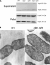
FIG 1
FIG 1
T3SS function and transmission electron microscopy. WT CO92 and the Δlpp, Δpla, and Δlpp Δpla mutants were grown overnight and then diluted 1:20 with fresh HIB. The cultures were grown for an additional 3 h at 28°C and then shifted to 37°C for 2 h. The secretion of YopE and YopH was induced by the addition of 5 mM EGTA. Culture supernatants and pellets were collected 5 min after EGTA addition. (A) The presence of YopE and YopH in the samples was analyzed by the use of antibodies to YopE and YopH. To evaluate membrane integrity, both WT CO92 and the Δlpp Δpla double mutant were grown to exponential phase at 28°C (OD600, 0.8). The cells were washed, pelleted, fixed, and subjected to transmission electron microscopy (B).
FIG 2
FIG 2
Intracellular survival of WT Y. pestis CO92 and its mutant strains in murine macrophages. RAW 264.7 macrophages were infected with the WT and its tested mutants at an MOI of 1 for 30 min. Monolayers were treated with 100 μg/ml gentamicin for 1 h to kill extracellular bacteria. At 4 h (after gentamicin treatment), macrophages were lysed and cultured on SBA plates. Three independent experiments were performed. The data were analyzed using one-way ANOVA with the Bonferroni correction, and a P value of ≤0.05 was considered significant. Asterisks, significant difference by comparison of the results for the mutants with those for WT CO92.
FIG 3
FIG 3
Survival analysis of mice infected with WT Y. pestis CO92 and its mutant strains by the route causing bubonic plague. Mice were challenged by the s.c. route with 5 × 10⁵ CFU (representing 10,000 LD50s of WT CO92) of WT Y. pestis CO92 and its various mutants at day 0 and observed for mortality. At 30 days p.i., mice that had survived the initial infection were challenged with 10 LD50s of WT CO92 by the i.n. route. We found a 90% survival rate for the Δlpp Δpla double mutant-immunized animals and a 77% survival rate for the Δpla single mutant-immunized mice following challenge with WT CO92. The data were analyzed by using Kaplan-Meier survival estimates, and P values of ≤0.05 were considered significant.
FIG 4
FIG 4
Dissemination of WT Y. pestis CO92 and its mutant strains to the peripheral organs of mice infected by the route causing bubonic plague. Mice (20 to 25 per group) were challenged by the s.c. route with 5 × 10⁵ CFU of WT Y. pestis CO92 and its various mutants at day 0. On days 2, 4, 8, and 16, five to seven animals per group were sacrificed and the spleen, liver, lungs, and blood were collected. Each organ (except for blood) was homogenized and plated to determine the bacterial load. The data were analyzed by one-way ANOVA and Tukey's post hoc test, and a P value of ≤0.05 was considered significant. Because of the terminal nature of some animals, blood could not be drawn from them.
FIG 5
FIG 5
Antibody responses of mice challenged with the mutant strains of Y. pestis CO92 by the route causing bubonic plague. Mice were challenged by the s.c. route with 5 × 10⁵ CFU of the Δpla single mutant or the Δlpp Δpla double mutant at day 0, and survivors were bled 16 days later to determine antibody titers. (A) To observe the general antibody responses (IgG) to Y. pestis, we grew WT Y. pestis CO92 at 28°C overnight and then shifted the temperature to 37°C for 4 h and used the bacteria to coat the plates for ELISA. An ELISA was also performed on plates coated with the F1-V antigens to measure the overall antigen-specific IgG response (B) and the response to the IgG1(C), IgG2a (D), and IgG2b (E) isotypes. The geometric mean of each sample (n = 5) was used for data plotting. The data were analyzed statistically by one-way ANOVA with Tukey's post hoc test, and P values of ≤0.05 were considered significant.
FIG 6
FIG 6
Survival analysis of mice infected with WT Y. pestis CO92 and its mutant strains by the route causing pneumonic plague. Mice (20 to 30 per group) were challenged by the i.n. route with 5 × 10⁵ CFU (representing 1,000 LD50s of WT CO92) of WT Y. pestis CO92 and its various mutants at day 0 and observed for mortality. At 30 days p.i., survivors were rechallenged with 10 LD50s of the WT CO92 strain by the i.n. route and observed for mortality. The data were statistically analyzed by using Kaplan-Meier's survival estimates, and a P value of ≤0.05 was considered significant.
FIG 7
FIG 7
Dissemination of WT Y. pestis CO92 and its mutants to peripheral organs of mice infected by the route causing pneumonic plague. Mice were challenged by the i.n. route with 5 × 10⁵ CFU of WT Y. pestis CO92 and its various mutants at day 0. On days 2, 6, 8, and 14, 5 to 10 animals per group were sacrificed and the spleen, liver, lungs, and blood were collected. Each organ (except for blood) was homogenized and plated to determine the bacterial load. The data were analyzed by one-way ANOVA and Tukey's post hoc test. Statistically significant values are indicated (***, P < 0.001), and these data were compared to those for animals infected with WT CO92. By day 2, all of the WT bacterium- and Δlpp mutant-infected mice had died. Because of the terminal nature of some animals, blood could not be drawn from them.
FIG 8
FIG 8
Antibody responses of mice challenged with the mutant strains of Y. pestis CO92 by the route causing pneumonic plague. Mice (n = 10) were challenged by the i.n. route with 5 × 10⁵ CFU of the Δpla single mutant or the Δlpp Δpla double mutant on day 0 and bled 14 days later to determine the antibody titers in serum. (A) WT Y. pestis CO92 was grown to coat the plates for ELISA to observe the total IgG antibody response to Y. pestis. An ELISA was also performed to examine the total antigen-specific IgG (B), IgG1 (C), IgG2a (D), and IgG2b (E) responses when the plates were coated with the F1-V antigens of Y. pestis. The geometric mean of each sample (n = 5) was used for plotting of the data. The data were analyzed statistically by one-way ANOVA with Tukey's post hoc test, and P values of ≤0.05 were considered significant.
FIG 9
FIG 9
Survival analysis of mice infected with WT Y. pestis CO92 and its complemented mutant strains by the route causing pneumonic plague. Mice (10 per group) were challenged by the i.n. route with 5 × 10⁵ CFU (representing 1,000 LD50s of WT CO92) of WT Y. pestis CO92, its various mutants, and the complemented Δpla single mutant or the Δlpp Δpla double mutant (at day 0) and observed for mortality. The data were analyzed by using Kaplan-Meier survival estimates, and a P value of ≤0.05 was considered significant. The data between WT CO92 and its various mutants as well as between the Δpla single mutant and the Δlpp Δpla double mutant were compared.
FIG 10
FIG 10
Histopathology of mouse tissues following infection with WT Y. pestis CO92 and mutant strains. Mice were challenged by the i.n. route with 5 × 10⁵ CFU of either WT CO92 or its mutants. At 2 days p.i., a portion of the lungs, liver, and spleen was stained with H&E and evaluated. Organs from 3 animals were examined, and representative data are shown here. Arrows in lung and liver images, inflammation; plus signs in lung images, observable bacterial presence; diamonds, edematous areas; asterisks in spleen images, areas of decreased cellularity in the red pulp and marginal zones of the white pulp.
FIG 11
FIG 11
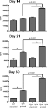
Proliferation of T cells isolated from WT Y. pestis CO92- and Δlpp Δpla mutant-infected mice. A total of 60 mice were i.n. challenged with a sublethal dose (250 CFU) of WT CO92 or the Δlpp Δpla double mutant (30 mice per group). At 14, 21, and 60 days p.i., T cells were isolated separately from the spleens of 5 mice in each infected group and stimulated for 3 days with APCs that had been pulsed with heat-killed WT CO92 and then γ-irradiated. Data were analyzed by using the Bonferroni correction with a one-way ANOVA, and a P value of ≤0.05 was considered significant. *, statistical significance in comparing unpulsed to pulsed T cells (P < 0.001) for both WT- and mutant-infected mice. The results represent averages from 2 independent experiments.
FIG 12
FIG 12
Cytokine production by T cells isolated from WT Y. pestis CO92- and CO92 mutant-immunized mice after ex vivo pulsing with plague bacterium antigens. A total of 60 mice were i.n. challenged with a sublethal dose (250 CFU) of the WT or the Δlpp Δpla mutant (30 mice per group). At 14, 21, and 60 days p.i., spleens from 5 mice in each infected group (at each time point) were harvested, and T cells were isolated from each animal separately (including uninfected controls) and incubated with pulsed or unpulsed APCs. After 48 h of incubation, supernatants were harvested for analysis by use of a Milliplex kit. Data were analyzed by use of the Bonferroni correction with one-way ANOVA, and a P value of ≤0.05 was considered significant.
FIG 13
FIG 13
Flow cytometry of T cells isolated from WT Y. pestis CO92- and CO92 mutant-immunized mice after ex vivo pulsing with plague bacterium antigens. T cells from animals infected with WT CO92 or the Δlpp Δpla double mutant or uninfected mice (see the legend to Fig. 12) were cultured with pulsed APCs for 5 days. Cells were then stained and read on a flow cytometer (LSR II Fortessa), and the data were analyzed by using FACS Diva software. T cells were stained on days 14 (CD4+0 cells [A] and CD8+ cells [C]) and 60 (CD4+ cells [B] and CD8+ cells [D]). These data represent averages from two independent experiments. Data were analyzed by Tukey's post hoc test with one-way ANOVA, and a P value of ≤0.05 was considered significant. *, statistical significance (P < 0.05) between control mice and WT CO92-infected or Δlpp Δpla double mutant-infected mice.

Similar articles

Cited by

References

    1. Prentice MB, Rahalison L. 2007. Plague. Lancet 369:1196–1207. 10.1016/S0140-6736(07)60566-2 - DOI - PubMed
    1. Huang XZ, Nikolich MP, Lindler LE. 2006. Current trends in plague research: from genomics to virulence. Clin. Med. Res. 4:189–199. 10.3121/cmr.4.3.189 - DOI - PMC - PubMed
    1. Ligon BL. 2006. Plague: a review of its history and potential as a biological weapon. Semin. Pediatr. Infect. Dis. 17:161–170. 10.1053/j.spid.2006.07.002 - DOI - PubMed
    1. Perry RD, Fetherston JD. 1997. Yersinia pestis—etiologic agent of plague. Clin. Microbiol. Rev. 10:35–66 - PMC - PubMed
    1. World Health Organization. 2006. Interregional meeting on prevention and control of plague, Antanarivo, Madagascar, 1 to 11 April. World Health Organization, Geneva, Switzerland: http://www.who.int/csr/resources/publications/WHO_HSE_EPR_2008_3w.pdf

Publication types

MeSH terms