. 2014 Jun;82(6):2417-23.
doi: 10.1128/IAI.01678-13. Epub 2014 Mar 31.

Novel pneumocystis antigen discovery using fungal surface proteomics

Affiliations

Novel pneumocystis antigen discovery using fungal surface proteomics

Mingquan Zheng et al. Infect Immun. 2014 Jun.

Erratum in

  • Infect Immun. 2014 Aug;82(8):3513

Abstract

Pneumonia due to the fungus Pneumocystis jirovecii is a life-threatening infection that occurs in immunocompromised patients. The inability to culture the organism as well as the lack of an annotated genome has hindered antigen discovery that could be useful in developing novel vaccine- or antibody-based therapies as well as diagnostics for this infection. Here we report a novel method of surface proteomics analysis of Pneumocystis murina that reliably detected putative surface proteins that are conserved in Pneumocystis jirovecii. This technique identified novel CD4(+) T-cell epitopes as well as a novel B-cell epitope, Meu10, which encodes a glycosylphosphatidylinositol (GPI)-anchored protein thought to be involved in ascospore assembly. The described technique should facilitate the discovery of novel target proteins for diagnostics and therapeutics for Pneumocystis infection.


Figures
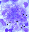
FIG 1
FIG 1
P. murina in BAL fluid of Pneumocystis-inoculated Rag2/Il2rg/ mice. Representative Diff-Quick staining of Pneumocystis in both cyst (denoted by arrows) and trophozoite (denoted by arrowheads) is shown.
FIG 2
FIG 2
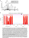
Identification of Pneumocystis surface peptides. (A) FPEK*IEVENLYK, an exposed portion of several P. murina major surface glycoproteins such as PENG_02417 and PNEG_00001. (B) The tandem mass spectrum of a doubly charged peptide precursor ion at m/z 925.1. HGQIEVTCAK*SGIYENSLWYIEDNS, a peptide expressed on the exposed portion of transmembrane P. murina gene PNEG_00837 (as predicted by TMHMM analysis), was searched against the P. murina protein sequence database using the in-house search engine Biomarks3.3. Modification of lysine (K*) by Sulfo-NHS-LC-biotin was unambiguously determined by the series of b and y ions on the tandem mass spectrum. The arrow indicates the site of labeling.
FIG 3
FIG 3
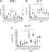
Response of CD4+ T-cell subsets to peptide stimulation. C57/B6 wild-type (wt) mice were inoculated with Pneumocystis for 2 weeks. Total lymphocytes from lung draining lymph node were stimulated by each peptide pool and control proteins in vitro for 4 days. Spot frequencies of IFN-γ, IL-5, and IL-17 were determined as spot-forming units/2 × 105 cells as plotted. All data are reported as means ± SEM for n = 4 per group. One-way ANOVA was applied with a Dunnett's multiple-comparison test. *, P < 0.05. Non Stl., nonstimulated; s. cer., S. cerevisiae; s. pom., S. pombe; PC Ag, Pneumocystis antigen.
FIG 4
FIG 4
Meu10 is an extracellular antigen capable of inducing a humoral immune response. (A) Recombinant myc-tagged Meu10 protein identified by SDS-PAGE and Western blotting in transfected 293 cell lysate. (B) Anti-Pneumocystis (PC) serum recognizes Meu10 lysate by ELISA compared to naive serum (top panel, P = 0.0009). Further normalization of the data to nontransfected 293 lysate (background) demonstrates that anti-Pneumocystis serum recognizes Meu10 at a greater optical density (O.D.) than naive serum (bottom panel, P = 0.024). (C) Immunofluorescent staining of whole Pneumocystis with anti-Meu10 serum binds to both the Pneumocystis cyst (top row) and the troph (middle row). Naive serum (bottom row) demonstrated very little specific binding. Anti-Meu10 (green) staining only is shown in the left column, while DAPI (blue) staining is shown in the middle column. The overlay of the two images, demonstrating Meu10 staining both cysts and trophs, is shown in the right column.

References

    1. Zheng M, Shellito JE, Marrero L, Zhong Q, Julian S, Ye P, Wallace V, Schwarzenberger P, Kolls JK. 2001. CD4+ T cell-independent vaccination against Pneumocystis carinii in mice. J. Clin. Invest. 108:1469–1474. 10.1172/JCI13826 - DOI - PMC - PubMed
    1. Zheng M, Ramsay AJ, Robichaux MB, Norris KA, Kliment C, Crowe C, Rapaka RR, Steele C, McAllister F, Shellito JE, Marrero L, Schwarzenberger P, Zhong Q, Kolls JK. 2005. CD4+ T cell-independent DNA vaccination against opportunistic infections. J. Clin. Invest. 115:3536–3544. 10.1172/JCI26306 - DOI - PMC - PubMed
    1. Empey KM, Hollifield M, Schuer K, Gigliotti F, Garvy BA. 2004. Passive immunization of neonatal mice against Pneumocystis carinii f. sp. muris enhances control of infection without stimulating inflammation. Infect. Immun. 72:6211–6220. 10.1128/IAI.72.11.6211-6220.2004 - DOI - PMC - PubMed
    1. Wells J, Haidaris CG, Wright TW, Gigliotti F. 2006. Active immunization against Pneumocystis carinii with a recombinant P. carinii antigen. Infect. Immun. 74:2446–2448. 10.1128/IAI.74.4.2446-2448.2006 - DOI - PMC - PubMed
    1. Gigliotti F, Haidaris CG, Wright TW, Harmsen AG. 2002. Passive intranasal monoclonal antibody prophylaxis against murine Pneumocystis carinii pneumonia. Infect. Immun. 70:1069–1074. 10.1128/IAI.70.3.1069-1074.2002 - DOI - PMC - PubMed

Publication types

MeSH terms