. 2015 Mar;20(3):361-8.
doi: 10.1038/mp.2014.22. Epub 2014 Apr 1.

Phenotypic differences in hiPSC NPCs derived from patients with schizophrenia

Affiliations

Phenotypic differences in hiPSC NPCs derived from patients with schizophrenia

K Brennand et al. Mol Psychiatry. 2015 Mar.

Abstract

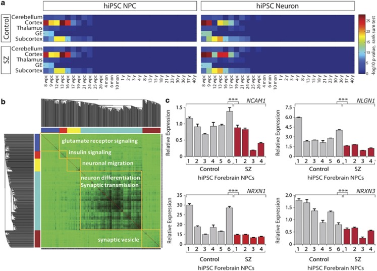
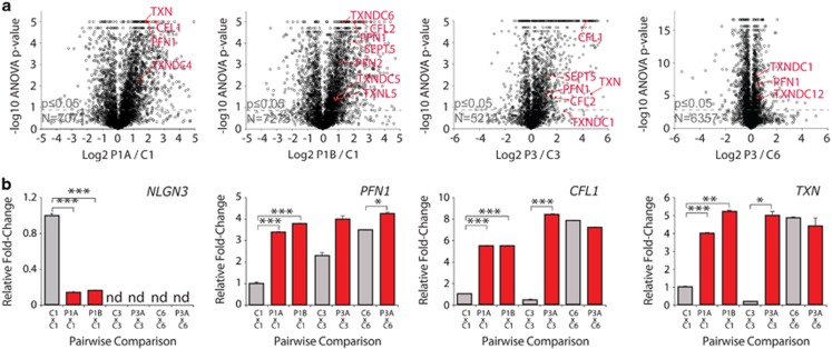
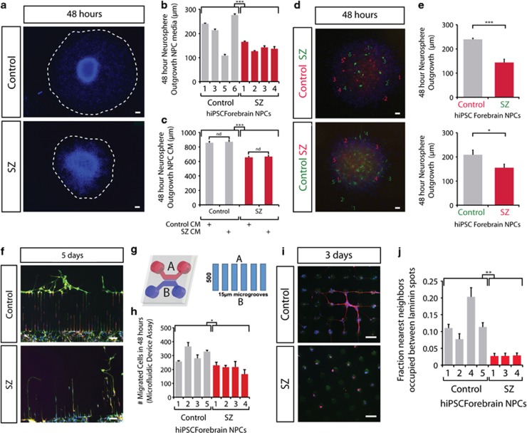
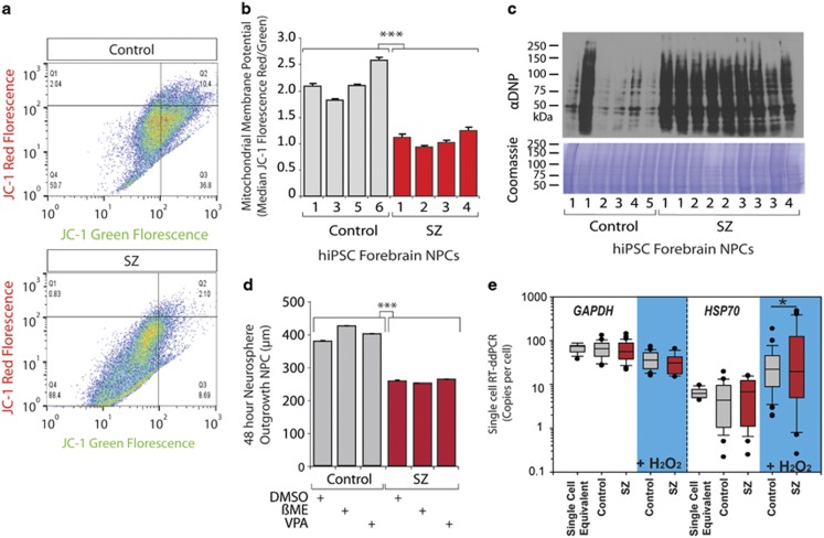
Consistent with recent reports indicating that neurons differentiated in vitro from human-induced pluripotent stem cells (hiPSCs) are immature relative to those in the human brain, gene expression comparisons of our hiPSC-derived neurons to the Allen BrainSpan Atlas indicate that they most resemble fetal brain tissue. This finding suggests that, rather than modeling the late features of schizophrenia (SZ), hiPSC-based models may be better suited for the study of disease predisposition. We now report that a significant fraction of the gene signature of SZ hiPSC-derived neurons is conserved in SZ hiPSC neural progenitor cells (NPCs). We used two independent discovery-based approaches-microarray gene expression and stable isotope labeling by amino acids in cell culture (SILAC) quantitative proteomic mass spectrometry analyses-to identify cellular phenotypes in SZ hiPSC NPCs from four SZ patients. From our findings that SZ hiPSC NPCs show abnormal gene expression and protein levels related to cytoskeletal remodeling and oxidative stress, we predicted, and subsequently observed, aberrant migration and increased oxidative stress in SZ hiPSC NPCs. These reproducible NPC phenotypes were identified through scalable assays that can be applied to expanded cohorts of SZ patients, making them a potentially valuable tool with which to study the developmental mechanisms contributing to SZ.


Figures

Figure 1
Figure 1
Gene expression of control and schizophrenia (SZ) human-induced pluripotent stem cell (hiPSC) neural progenitor cells (NPCs) and 6-week-old neurons most resembles human first trimester forebrain tissue. (a) Heatmaps produced by Wilcoxon's rank-sum comparisons of control and SZ hiPSC forebrain NPC and 6-week-old neuron microarray gene expression relative to the Allen BrainSpan Atlas. (b) WGCNA (weighted gene co-expression network analysis) of the SZ NPC gene signature identified five modules. (c) Quantitative PCR validation of the altered expression of the adhesion genes NCAM1, NLGN1, NRXN1 and NRXN3 in hiPSC forebrain NPCs from six controls and four SZ patients. Error bars are s.e.m., ***P<0.001. See also Supplementary Figures 1–6.
Figure 2
Figure 2
Aberrant expression of cytoskeletal and oxidative stress proteins in four independent pairwise SILAC (stable isotope labeling by amino acids in cell culture) quantitative proteomic mass spectrometry comparisons of control and schizophrenia (SZ) human induced pluripotent stem cell (hiPSC) forebrain neural progenitor cells (NPCs). (a) Volcano plots of −log10 analysis of variance P-value versus log2 SZ/control protein levels for pairwise analyses of NPCs from patient 1 (left) and patient 3 (right) compared with gender-matched controls 1, 3 or 6, respectively. Key cytoskeletal remodeling proteins (cofilins and profilins) and oxidative stress proteins (thioredoxin and related proteins) are highlighted. From left to right, N=7072, 7226, 5215 and 6357 proteins. (b) Bar graphs showing decreased NLGN3 in P1 SILAC comparisons and increased PFN1, CFL1 and TXN protein levels in four independent SILAC comparisons. Error bars are s.e.m., *P<0.05, **P<0.01, ***P<0.001. See also Supplementary Figure 5.
Figure 3
Figure 3
Aberrant migration in schizophrenia (SZ) human-induced pluripotent stem cell (hiPSC) forebrain neural progenitor cells (NPCs). (a) Representative images of hiPSC forebrain NPC neurosphere outgrowth assay. The average distance between the radius of the inner neurosphere (dense aggregate of nuclei) and outer circumference of cells (white dashed line) was calculated. 4′,6-Diamidino-2-phenylindole (DAPI)-stained nuclei (blue). Scale bar, 100 μm. (b) Neurosphere outgrowth by control and SZ hiPSC forebrain NPCs. (c) Neurosphere outgrowth following coculture with control or SZ hiPSC NPC-conditioned media (CM). (d) Representative images of mixed control and SZ hiPSC NPCs, labeled with lentivirus-green fluorescent protein and lentivirus-red fluorescent protein. Green and red numbers indicate five furthest migrated lentiviral-green fluorescent protein and lentiviral-red fluorescent protein NPCs in reciprocal migration experiments. DAPI-stained nuclei (blue). Scale bar 100 μm. (e) Neurosphere outgrowth in neurospheres composed of mixed control and SZ hiPSC NPCs. (f) Representative images of hiPSC forebrain NPCs after 5 days in a microfluidic device. At time 0, all NPCs were below the microgrooves pictured—migration occurred up from chamber B into chamber A. Migrating neural cells stained with MAP2AB (red), βIII-tubulin (green); DAPI-stained nuclei (blue). Scale bar, 100 μm. (g) Schematic of the microfluidic chambers. At time 0, hiPSC NPCs were added to chamber B and allowed to begin migration toward chamber A via 15-μm grooves. (h) Cellular migration of control and SZ hiPSC NPCs in microfluidic devices at 48 h. (i) Representative images of hiPSC forebrain NPCs in micropatterned laminin spot migration assay. Laminin spot stained (green); migrating neural cells stained with βIII-tubulin (red); and DAPI-stained nuclei (blue). Scale bar, 100 μm. (j) Nearest-neighbor analysis of ‘chaining' between laminin spots by control and SZ hiPSC forebrain NPCs. Error bars are s.e.m., *P<0.05, **P<0.01, ***P<0.001. See also Supplementary Figures 6–8.
Figure 4
Figure 4
Mitochondrial damage and increased oxidative stress in schizophrenia (SZ) human-induced pluripotent stem cell (hiPSC) neural progenitor cells (NPCs). (a) Representative fluorescence-activated cell sorting (FACS) plots for JC-1 red/green fluorescence in control and SZ hiPSC NPCs. (b) FACS analysis for mitochondrial membrane potential (MMP) in control and SZ hiPSC NPCs indicated by median JC-1 red/green fluorescence. (c) OxyBlot western blot for oxidized proteins in SZ hiPSC NPCs. (d) Neurosphere outgrowth by control and SZ hiPSC forebrain NPCs with and without treatment by the anti-oxidant β-mercaptoethanol or valproic acid (VPA). (e) Graph shows fold change of GAPDH and HSP70 expression following H2O2 treatment compared with PBS exposure. There was significantly increased variability observed in SZ hiPSC NPCs as compared with the control (n>50), but no significant differences in the means in all sets of comparisons. Error bars are s.e.m., *P<0.05, ***P<0.001. See also Supplementary Figure 9.

References

    1. Weinberger DR. Implications of normal brain development for the pathogenesis of schizophrenia. Arch Gen Psychiatry. 1987;44:660–669. - PubMed
    1. White T, Anjum A, Schulz SC. The schizophrenia prodrome. Am J Psychiatry. 2006;163:376–380. - PubMed
    1. Wong AH, Van Tol HH. Schizophrenia: from phenomenology to neurobiology. Neurosci Biobehav Rev. 2003;27:269–306. - PubMed
    1. Gulsuner S, Walsh T, Watts AC, Lee MK, Thornton AM, Casadei S, et al. Spatial and temporal mapping of de novo mutations in schizophrenia to a fetal prefrontal cortical network. Cell. 2013;154:518–529. - PMC - PubMed
    1. Nicholas CR, Chen J, Tang Y, Southwell DG, Chalmers N, Vogt D, et al. Functional maturation of hPSC-derived forebrain interneurons requires an extended timeline and mimics human neural development. Cell Stem Cell. 2013;12:573–586. - PMC - PubMed

Publication types

MeSH terms

Substances