. 2014 May 15;74(10):2688-97.
doi: 10.1158/0008-5472.CAN-13-2582. Epub 2014 Mar 31.

Distinguishing between benign and malignant melanocytic nevi by in vivo multiphoton microscopy

Affiliations

Distinguishing between benign and malignant melanocytic nevi by in vivo multiphoton microscopy

Mihaela Balu et al. Cancer Res. .

Abstract

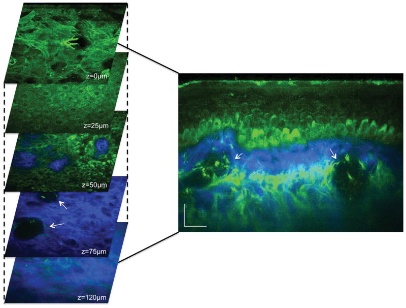
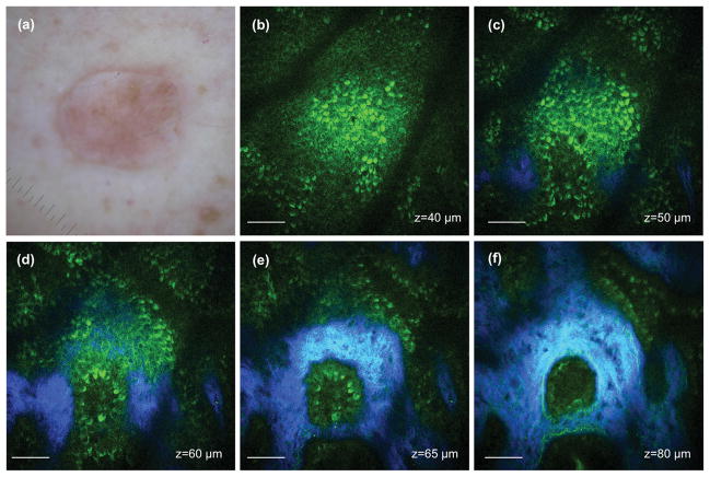
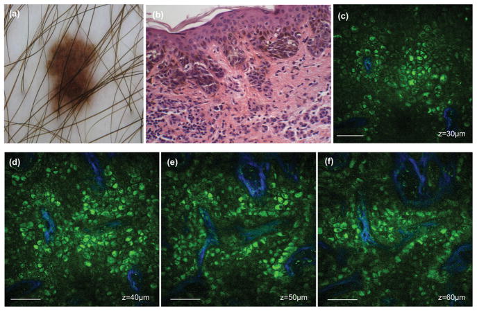
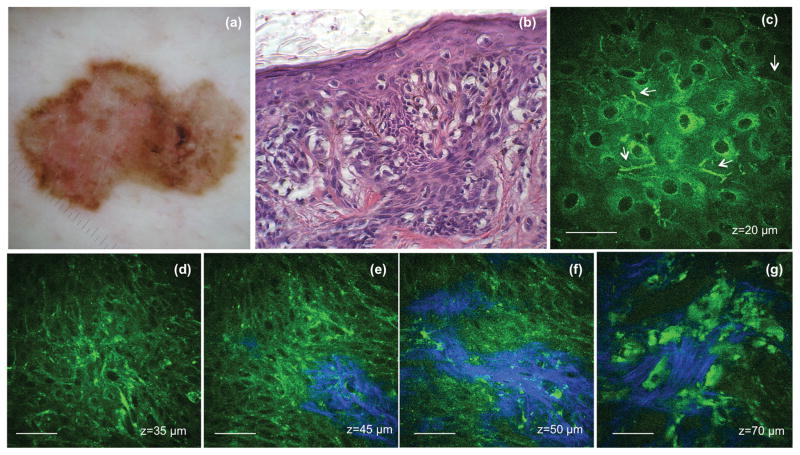
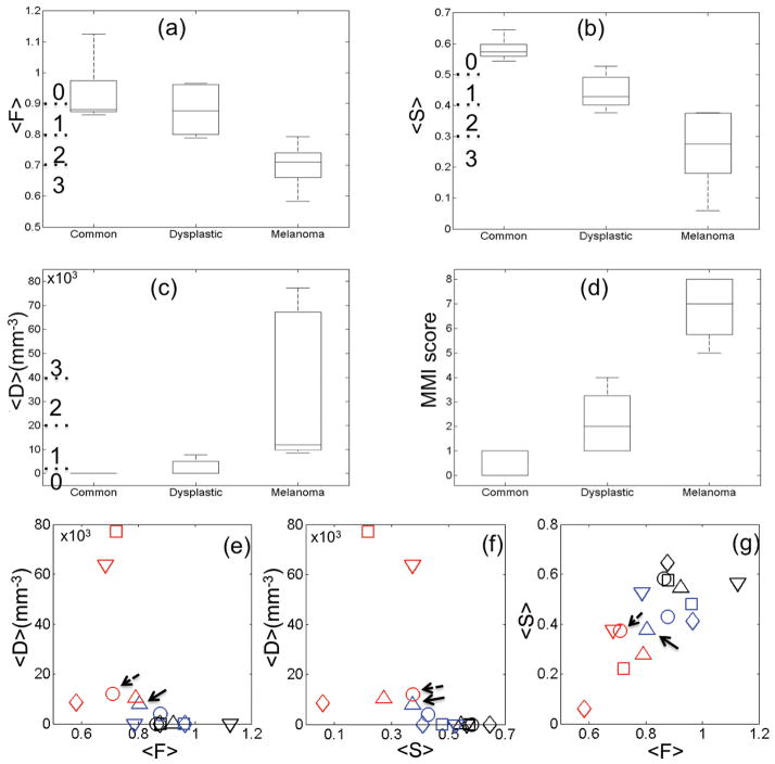
Monitoring of atypical nevi is an important step in early detection of melanoma, a clinical imperative in preventing the disease progression. Current standard diagnosis is based on biopsy and histopathologic examination, a method that is invasive and highly dependent upon physician experience. In this work, we used a clinical multiphoton microscope to image in vivo and noninvasively melanocytic nevi at three different stages: common nevi without dysplastic changes, dysplastic nevi with structural and architectural atypia, and melanoma. We analyzed multiphoton microscopy (MPM) images corresponding to 15 lesions (five in each group) both qualitatively and quantitatively. For the qualitative analysis, we identified the morphologic features characteristic of each group. MPM images corresponding to dysplastic nevi and melanoma were compared with standard histopathology to determine correlations between tissue constituents and morphology and to evaluate whether standard histopathology criteria can be identified in the MPM images. Prominent qualitative correlations included the morphology of epidermal keratinocytes, the appearance of nests of nevus cells surrounded by collagen fibers, and the structure of the epidermal-dermal junction. For the quantitative analysis, we defined a numerical multiphoton melanoma index (MMI) based on three-dimensional in vivo image analysis that scores signals derived from two-photon excited fluorescence, second harmonic generation, and melanocyte morphology features on a continuous 9-point scale. Indices corresponding to common nevi (0-1), dysplastic nevi (1-4), and melanoma (5-8) were significantly different (P < 0.05), suggesting the potential of the method to distinguish between melanocytic nevi in vivo.


Conflict of interest statement

Conflict of interest

Karsten König is cofounder of JenLab GmbH.

Figures

Figure 1
Figure 1. Pigmented normal skin
(Left) Horizontal sections of MPM images (XY scans) at different depths showing images of the stratum corneum (z=0μm), keratinocytes normally distributed in the stratum spinosum (z=25μm), the basal cells (green) surrounding dermal papilla (blue) (z=50μm), collagen fibers (blue) and cross-sections of blood vessels (white arrows) (z=75μm), collagen (blue) and elastin (green) in the dermis (z=120μm). (Right) Cross-sectional view (XZ scan) corresponding to a vertical plane through the horizontal sections on the left. Arrows point to blood vessels.
Figure 2
Figure 2. Compound nevus
(a) Clinical image (DermLite FOTO, Dermlite Inc.) (b) MPM image of the basal layer showing nevus cells and pigmented basal cells; F=1.02 (c) MPM image of the basal layer showing nevus cells, pigmented basal cells (green) and appearance of dermal papilla (blue); F=1.04 (d) MPM images showing a nest of nevus cells (green) surrounded by collagen (blue) fibers; F=0.95 (e–f) MPM images at different depths showing a nest of nevus cells (green) surrounded by collagen (blue) and elastin (green) fibers. The S value for this stack is 0.51. Scale bar is 40 μm.
Figure 3
Figure 3. Dysplastic nevus
Clinical image (DermLite FOTO, Dermlite Inc.) (a) Histologic section of the lesion (b) MPM images showing irregular nests of nevus cells (green) and collagen fibers (blue) along the basal layer at depths of 30 μm (c), 40 μm (d), 50 μm (e) and 60 μm (f). The corresponding F values are 0.89 (c) and 0.86 (d). The S value for this stack is 0.41. Scale bar is 40 μm.
Figure 4
Figure 4. Melanoma - superficial spreading type
(a) Clinical image (DermLite FOTO, Dermlite Inc.) (b) Histologic section of the lesion (c) MPM images showing ascending melanocytes (arrows) in the granulosum layer of the epidermis (d) MPM images of the basal layer showing proliferation of atypical melanocytes (highly pleomorphic melanocytes) F=0.69 (e–f) MPM images of the basal layer showing basal cells and atypical melanocytes (green) surrounding dermal papilla (blue); F=0.63 and 0.62 respectively (g) MPM images showing melanoma cells and probably melanophages invading the dermis (blue-collagen fibers). The S value for this stack is 0.35. Scale bar is 40 μm.
Figure 5
Figure 5. Distribution and correlation of the mean values and scores
The distribution of the mean values of F (a), S (b) and D (c) parameters for common nevi, dysplastic nevi and melanoma. (d) The distribution of the MMI scores for common nevi, dysplastic nevi and melanoma. (e–g) Correlation of F, S and D values. The combination (color, marker) corresponds to an individual lesion. Black, blue and red represent the ‘common nevi’, the ‘dysplastic nevi’ and the ‘melanoma’ groups, respectively. Arrows point to the markers corresponding to the lesions discussed in the text.

References

    1. Siegel R, Naishadham D, Jemal A. Cancer statistics, 2012. CA Cancer J Clin. 2012;62:10–29. - PubMed
    1. Howlader N, Noone A, Krapcho M, Neyman N, Aminou R, Waldron W, et al. SEER Cancer Statistics Review, 1975–2008. National Cancer Institute; Bethesda, MD: 2011. http://seer.cancer.gov/csr/1975_2008/, based on November 2010 SEER data submission, posted to the SEER web site, 2011.
    1. Balch CM, Gershenwald JE, Soong SJ, Thompson JF, Atkins MB, Byrd DR, et al. Final version of 2009 AJCC melanoma staging and classification. Journal of clinical oncology: official journal of the American Society of Clinical Oncology. 2009;27:6199–206. - PMC - PubMed
    1. Abbasi NR, Shaw HM, Rigel DS, Friedman RJ, McCarthy WH, Osman I, et al. Early diagnosis of cutaneous melanoma - Revisiting the ABCD criteria. Jama-J Am Med Assoc. 2004;292:2771–6. - PubMed
    1. Osborne JE, Bourke JF, Graham-Brown RA, Hutchinson PE. False negative clinical diagnoses of malignant melanoma. Br J Dermatol. 1999;140:902–8. - PubMed

Publication types

MeSH terms