

E3 ubiquitin ligase HOIP attenuates apoptotic cell death induced by cisplatin
- PMID: 24686174
- PMCID: PMC3990471
- DOI: 10.1158/0008-5472.CAN-13-2131
E3 ubiquitin ligase HOIP attenuates apoptotic cell death induced by cisplatin
Abstract
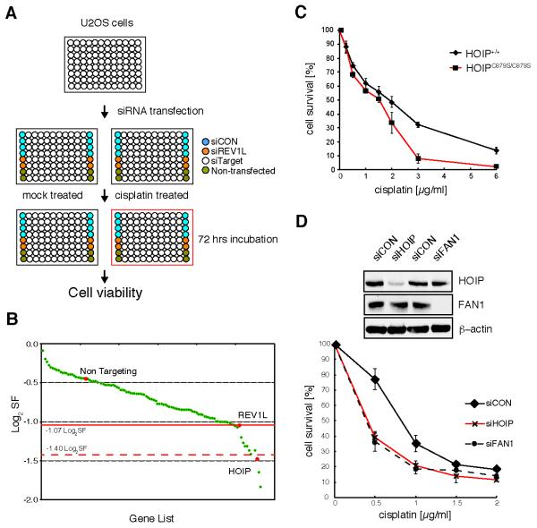
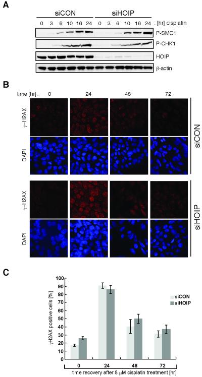
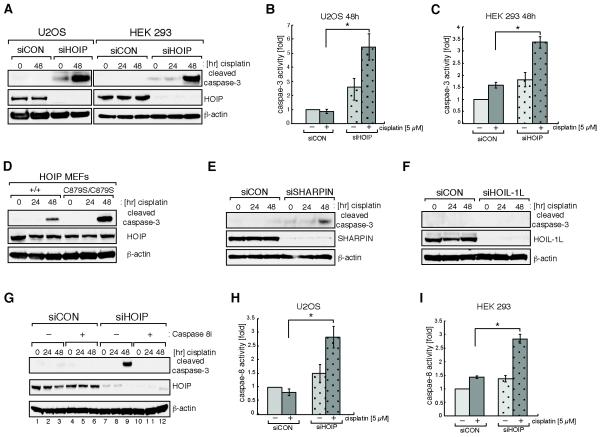
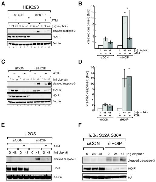
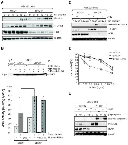
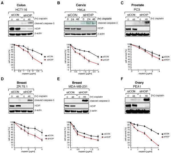
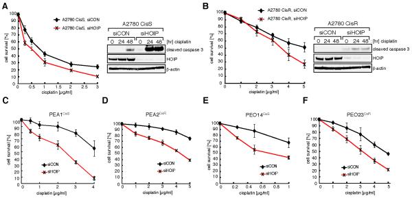
The genotoxin cisplatin is commonly used in chemotherapy to treat solid tumors, yet our understanding of the mechanism underlying the drug response is limited. In a focused siRNA screen, using an siRNA library targeting genes involved in ubiquitin and ubiquitin-like signaling, we identified the E3 ubiquitin ligase HOIP as a key regulator of cisplatin-induced genotoxicity. HOIP forms, with SHARPIN and HOIL-1L, the linear ubiquitin assembly complex (LUBAC). We show that cells deficient in the HOIP ligase complex exhibit hypersensitivity to cisplatin. This is due to a dramatic increase in caspase-8/caspase-3-mediated apoptosis that is strictly dependent on ATM-, but not ATR-mediated DNA damage checkpoint activation. Moreover, basal and cisplatin-induced activity of the stress response kinase JNK is enhanced in HOIP-depleted cells and, conversely, JNK inhibition can increase cellular resistance to cisplatin and reverse the apoptotic hyperactivation in HOIP-depleted cells. Furthermore, we show that HOIP depletion sensitizes cancer cells, derived from carcinomas of various origins, through an enhanced apoptotic cell death response. We also provide evidence that ovarian cancer cells classified as cisplatin-resistant can regain sensitivity following HOIP downregulation. Cumulatively, our study identifies a HOIP-regulated antiapoptotic signaling pathway, and we envisage HOIP as a potential target for the development of combinatorial chemotherapies to potentiate the efficacy of platinum-based anticancer drugs.
©2014 AACR.
Figures
Similar articles
-
A novel HOIP frameshift variant alleviates NF-kappaB signalling and sensitizes cells to TNF-induced death.Biochim Biophys Acta Mol Basis Dis. 2024 Oct;1870(7):167355. doi: 10.1016/j.bbadis.2024.167355. Epub 2024 Jul 14. Biochim Biophys Acta Mol Basis Dis. 2024. PMID: 39009172
-
Mechanistic insights into the subversion of the linear ubiquitin chain assembly complex by the E3 ligase IpaH1.4 of Shigella flexneri.Proc Natl Acad Sci U S A. 2022 Mar 22;119(12):e2116776119. doi: 10.1073/pnas.2116776119. Epub 2022 Mar 16. Proc Natl Acad Sci U S A. 2022. PMID: 35294289 Free PMC article.
-
CRL4 ubiquitin ligase stimulates Fanconi anemia pathway-induced single-stranded DNA-RPA signaling.BMC Cancer. 2019 Nov 5;19(1):1042. doi: 10.1186/s12885-019-6305-x. BMC Cancer. 2019. PMID: 31690264 Free PMC article.
-
Biochemistry, Pathophysiology, and Regulation of Linear Ubiquitination: Intricate Regulation by Coordinated Functions of the Associated Ligase and Deubiquitinase.Cells. 2021 Oct 9;10(10):2706. doi: 10.3390/cells10102706. Cells. 2021. PMID: 34685685 Free PMC article. Review.
-
Linear ubiquitin chains: enzymes, mechanisms and biology.Open Biol. 2017 Apr;7(4):170026. doi: 10.1098/rsob.170026. Open Biol. 2017. PMID: 28446710 Free PMC article. Review.
Cited by
-
Linear ubiquitin chains: NF-κB signalling, cell death and beyond.Nat Rev Mol Cell Biol. 2014 Aug;15(8):503-8. doi: 10.1038/nrm3836. Epub 2014 Jul 9. Nat Rev Mol Cell Biol. 2014. PMID: 25027653 Review.
-
Understanding and Targeting Apoptotic Pathways in Ovarian Cancer.Cancers (Basel). 2019 Oct 24;11(11):1631. doi: 10.3390/cancers11111631. Cancers (Basel). 2019. PMID: 31652965 Free PMC article. Review.
-
SHARPIN-mediated regulation of protein arginine methyltransferase 5 controls melanoma growth.J Clin Invest. 2018 Jan 2;128(1):517-530. doi: 10.1172/JCI95410. Epub 2017 Dec 11. J Clin Invest. 2018. PMID: 29227283 Free PMC article.
-
The MALDI-TOF E2/E3 Ligase Assay as Universal Tool for Drug Discovery in the Ubiquitin Pathway.Cell Chem Biol. 2018 Sep 20;25(9):1117-1127.e4. doi: 10.1016/j.chembiol.2018.06.004. Epub 2018 Jul 12. Cell Chem Biol. 2018. PMID: 30017913 Free PMC article.
-
Phenotypic screening using large-scale genomic libraries to identify drug targets for the treatment of cancer.Oncol Lett. 2020 Jun;19(6):3617-3626. doi: 10.3892/ol.2020.11512. Epub 2020 Apr 3. Oncol Lett. 2020. PMID: 32391087 Free PMC article. Review.
References
-
- Kelland L. The resurgence of platinum-based cancer chemotherapy. Nat Rev Cancer. 2007;7:573–84. - PubMed
-
- Koberle B, Tomicic MT, Usanova S, Kaina B. Cisplatin resistance: preclinical findings and clinical implications. Biochimica et biophysica acta. 2010;1806:172–82. - PubMed
-
- Holohan C, Van Schaeybroeck S, Longley DB, Johnston PG. Cancer drug resistance: an evolving paradigm. Nature reviews Cancer. 2013;13:714–26. - PubMed
-
- Ulrich HD, Walden H. Ubiquitin signalling in DNA replication and repair. Nat Rev Mol Cell Biol. 2010;11:479–89. - PubMed
Publication types
MeSH terms
Substances
Grants and funding
LinkOut - more resources
Full Text Sources
Other Literature Sources
Research Materials
Miscellaneous
