Review
. 2014 Aug;11(8):470-9.
doi: 10.1038/nrgastro.2014.46. Epub 2014 Apr 1.

Radiation enteropathy--pathogenesis, treatment and prevention

Affiliations
Review

Radiation enteropathy--pathogenesis, treatment and prevention

Martin Hauer-Jensen et al. Nat Rev Gastroenterol Hepatol. 2014 Aug.

Abstract

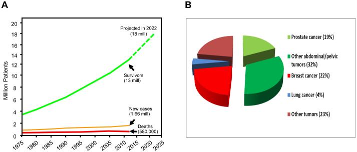
Changes in cancer incidence and mortality have been modest during the past several decades, but the number of cancer survivors has almost tripled during the same period. With an increasing cohort of cancer survivors, efforts to prevent, diagnose and manage adverse effects of cancer therapy, in general, and those of radiation therapy specifically, have intensified. Many cancer survivors have undergone radiation therapy of tumours in the pelvis or abdomen, thus rendering the bowel at risk of injury. In fact, the current prevalence of patients who have long-term radiation-induced intestinal adverse effects exceeds that of IBD. Considerable progress towards reducing toxicity of radiation therapy has been made by the introduction of so-called dose-sculpting treatment techniques, which enable precise delivery of the radiation beam. Moreover, new insights into the underlying pathophysiology have resulted in an improved understanding of mechanisms of radiation-induced bowel toxicity and in development of new diagnostic strategies and management opportunities. This Review discusses the pathogenesis of early and delayed radiation-induced bowel toxicity, presents current management options and outlines priorities for future research. By adding insight into molecular and cellular mechanisms of related bowel disorders, gastroenterologists can substantially strengthen these efforts.


Figures

Figure 1
Figure 1. Cancer survivors and cancer prevalence rates in the US
A) Cancer incidence and death rates have been fairly flat during the past 4 decades, while the cohort of cancer survivors increases by 3% per year, exceeds 13 million in 2013, and is expected to reach 18 million in 2022 . B) Approximately half of all cancer survivors are survivors after abdominal or pelvic tumors , many of whom have had or will have radiation therapy. This points to radiation enteropathy as a major obstacle to uncomplicated cancer cures.
Figure 2
Figure 2. Radiation mucositis (rat intestine, all images original magnification 40X)
A) Bioluminescence image (luminol) of an unirradiated (control) rat with intestine transposed to the left scrotum for fractionated irradiation. No increase in bioluminescence indicating little or no myeloperoxidase activity. B) Unirradiated (control) rat small intestine stained with anti-transferrin antibody. Few granulocytes are seen in the mucosa/submucosa. C) Unirradiated (control) rat small intestine stained with anti-ED2 (CD163) antibody. There are few macrophages in the mucosa/submucosa. D) Bioluminescence image (luminol) 5 days after localized irradiation of a segment of small bowel transposed to the left scrotum. Significant increase in bioluminescence indicating substantial myeloperoxidase activity. E) Intestine procured 2 weeks after irradiation showing accumulation of granulocytes in mucosa/submucosa. F) Intestine procured 2 weeks after irradition showing accumulation of macrophages in mucosa/submucosa.
Figure 3
Figure 3. Human endoscopic biopsies of rectal mucosa obtained from patient before and during ongoing radiation therapy of prostate cancer
A) Periodic acid Schiff (PAS)-staining of normal rectal mucosa before start of radiation therapy. Note intact surface epithelium, straight glands, and the many PAS-positive goblet cells (original magnification 20X). B) Glandular atrophy, mucosal inflammation, and loss of PAS-positive goblet cells 2 weeks into the course of radiation therapy (original magnification 20X).
Figure 4
Figure 4. Resection specimens of normal human small bowel and delayed radiation enteropathy
A) Normal intestine (original magnification 0.5X). B) Resected small intestine from a woman with severe delayed radiation enteropathy. Note atrophic mucosa and severe fibrosis in submucosa and subserosa (original magnification 0.5X, same as panel A).
Figure 5
Figure 5. Involvement of the intestinal immune system and microvascular endothelium in the regulation of acute radiation mucositis and subsequent adverse tissue remodeling (intestinal fibrosis)
When the mucosal barrier becomes disrupted, as after radiation exposure, bacterial products and other activating agents gain access to the intestinal tissues and stimulate a variety of immune cells to produce cytokines and other pro- and anti-inflammatory mediators. Moreover, radiation-induced endothelial dysfunction leads to endothelial dysfunction with loss of thromboresistance, resulting in thrombin formation, neutrophil recruitment and activation, and stimulation of mesenchymal cells. In addition to the mechanisms depicted here, a host of other mechanisms, for example, those related to mast cells, the enteric nervous system, and the gut microbiome plays important roles in the pathogenesis of radiation enteropathy (see the section Important Unanswered Questions in Radiation Enteropathy).
Figure 6
Figure 6. Radiation-induced changes in the proximal jejunum from the non-human primate
Proximal jejunum from unirradiated Rhesus macaque (A) and Rhesus macaque 4-12 days after exposure to single-dose irradiation (B-D). Note conspicuous disappearance of plicae circulares, crypt irregularity, and pronounced villus atrophy after irradiation. Partial recovery of post-irradiation changes is seen at 12 days, with near complete recovery of the epithelium and beginning re-appearance of plicae circulares. Original magnification of all images 1.4X.
Figure 7
Figure 7
Algorithm depicting simplified principles of work-up and common approaches for managing patients with delayed gastrointestinal symptoms after radiation therapy

References

    1. Goitein M. How best to dispose of extra-tumoral dose: a cautionary note for intensity-modulated radiation therapy. Int J Radiat Oncol Biol Phys. 2009;75:1–3. - PubMed
    1. Eifel PJ, Jhingran A, Bodurka DC, Levenback C, Thames H. Correlation of smoking history and other patient characteristics with major complications of pelvic radiation therapy for cervical cancer. J Clin Oncol. 2002;20:3651–3657. - PubMed
    1. Wedlake LJ, et al. Predicting late effects of pelvic radiotherapy: is there a better approach? Int J Radiat Oncol Biol Phys. 2010;78:1163–1170. - PubMed
    1. Willett CG, et al. Acute and late toxicity of patients with inflammatory bowel disease undergoing irradiation for abdominal and pelvic neoplasms. Int J Radiat Oncol Biol Phys. 2000;46:995–998. - PubMed
    1. Herold DM, Hanlon AL, Hanks GE. Diabetes mellitus: a predictor for late radiation morbidity. Int J Radiat Oncol Biol Phys. 1999;43:475–479. - PubMed

Publication types