. 2014 Jul:67:79-87.
doi: 10.1016/j.nbd.2014.03.011. Epub 2014 Mar 29.

High fat diet produces brain insulin resistance, synaptodendritic abnormalities and altered behavior in mice

Affiliations

High fat diet produces brain insulin resistance, synaptodendritic abnormalities and altered behavior in mice

Steven E Arnold et al. Neurobiol Dis. 2014 Jul.

Abstract

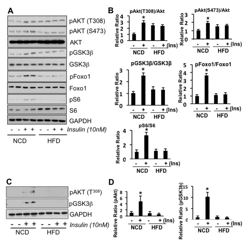
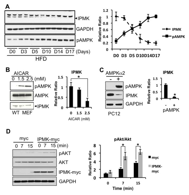
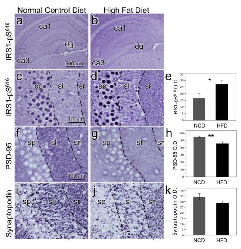
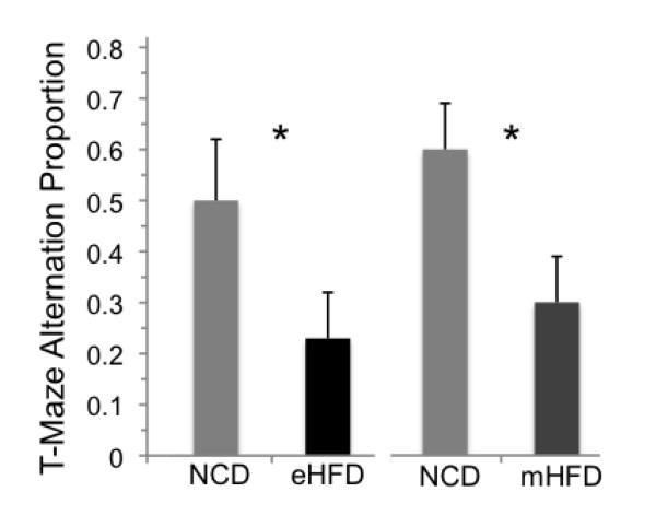
Insulin resistance and other features of the metabolic syndrome are increasingly recognized for their effects on cognitive health. To ascertain mechanisms by which this occurs, we fed mice a very high fat diet (60% kcal by fat) for 17days or a moderate high fat diet (HFD, 45% kcal by fat) for 8weeks and examined changes in brain insulin signaling responses, hippocampal synaptodendritic protein expression, and spatial working memory. Compared to normal control diet mice, cerebral cortex tissues of HFD mice were insulin-resistant as evidenced by failed activation of Akt, S6 and GSK3β with ex-vivo insulin stimulation. Importantly, we found that expression of brain IPMK, which is necessary for mTOR/Akt signaling, remained decreased in HFD mice upon activation of AMPK. HFD mouse hippocampus exhibited increased expression of serine-phosphorylated insulin receptor substrate 1 (IRS1-pS(616)), a marker of insulin resistance, as well as decreased expression of PSD-95, a scaffolding protein enriched in post-synaptic densities, and synaptopodin, an actin-associated protein enriched in spine apparatuses. Spatial working memory was impaired as assessed by decreased spontaneous alternation in a T-maze. These findings indicate that HFD is associated with telencephalic insulin resistance and deleterious effects on synaptic integrity and cognitive behaviors.

Keywords: Akt; GSK3; IPMK; Insulin; Insulin receptor substrate 1; Spine; Synapse; Working memory; mTOR.


Figures

Figure 1
Figure 1
Representative Western blots of frontal brain tissue from HFD or NCD-fed mice with (+) or without (-) ex vivo insulin stimulation. Mice (n=5-10) were fed with either a 60% HFD for 17 days or 45% HFD for 8 weeks. A) Representative blots from pooled brain tissues of 60% HFD or NCD mice show that insulin stimulation in HFD mouse brain tissue fails to activate IRS-1, Akt, GSK3β, S6 (downstream of mTOR) and Foxo1 (a direct downstream target of Akt). GAPDH served as a loading control. B) Western blot quantitation in 60% HFD vs NCD mice. C) Representative blots from tissue samples from 45% HFD or NCD mice shows HFD mouse brain failure to activate AKT and GSK3 with insulin stimulation. D) Western blot quantitation in 45% HFD vs NCD mice. Student’s t-tests were performed (* p < 0.05).
Figure 2
Figure 2
A potential interaction between IPMK and pAMPK. A) Mice fed a 60% HFD for 17 days show a gradual decrease in IPMK while the levels of pAMPK increase (means + SEM). B-C) When pAMPK is induced either by overexpression of catalytic subunit of AMPK in differentiated PC12 cells or by a pharmacological reagent, IPMK levels are decreased. D) Differentiated PC12 cells overexpressed with either myc or IPMK-myc were treated with 100nM insulin for various time points. Overexpression of IPMK further sensitizes insulin-mediated-activation of pAkt. (* p < 0.05)
Figure 3
Figure 3
HFD effects on hippocampal CA3 molecular neuroanatomy. Photomicorgraphs of hippocampus) from mice fed a 60% fat calorie HFD or NCD for 17 days. A) Low magnification image of hippocampus labeled for serine phosphorylated IRS1-pS616 with asterisk indicating location in CA3 where higher power image in C was captured. B) Low magnification image of HFD mouse with asterisk for image in D. C) CA3 stratum pyramidale (sp) immunolabeled IRS1-pS616 shows normal nuclear labeling and very little cytoplasmic membrane or neurite labeling in stratum lucidum (sl = mossy fiber terminal zone). D) HFD mouse CA3 shows increase in overall immunolabeling with abnormal neuritic labeling. E) NCD CA3 stratum lucidum with pseudocolored double immunofluorescence labeling for pS-IRS1 (red) and spinophilin (green) at high magnification. Spinophilin is a regulatory subunit of protein phosphatase-1 catalytic subunit that is highly enriched in dendritic spines. Note the virtual absence of pS-IRS1 labeling of this dendrite and spine-populated region. F) HFD mouse double immunolabeling shows increased pS-IRS1 dendritic labeling in red and decreased numbers of labeled spines in green.
Figure 4
Figure 4
Chronic moderate HFD effects on hippocampal CA3 molecular neuroanatomy. Photomicrographs are from mice fed a 45% fat calorie HFD or NCD for 8 weeks. A) Low magnification image of NCD mouse hippocampus labeled for IRS1-pS616 with box indicating location in CA3 where higher power images in C was taken. B) Low magnification image of 45% HFD mouse with box for image in D. C) NCD mouse CA3 labeled for IRS1-pS61 shows typical nuclear staining in stratum pyramidale (sp) and weak labeling of stratum lucidum (sl) and stratum radiatum (sr). D) 45% HFD mouse labeled for IRS1-pS616 shows increased overall labeling and distinctly labeled dendrites. E) Bar graph depicts mean optical density (O.D.) values (+SEM) of sl in NCD and 45% HFD mice (*p<0.05). F) NCD mouse CA3 labeled for PSD-95, an important scaffolding protein for post-synaptic densities. G) 45% HFD mouse CA3 shows decreased PSD-95 immunolabeling in sl. H) Graph depicting decreased PSD-95 expression in sl of HFD mice (**p<0.005). I) NCD mouse CA3 labeled for synaptopodin, an actin-associated protein essential for the formation of spine apparatuses in spines. Note its punctate appearance denoting spines. J) 45% HFD mouse CA3 shows decreased density of synaptopodin puncta in sl. K) Graph portrays decreased synaptopodin spine density in HFD mice (p<0.1).
Figure 5
Figure 5
Mice fed an extreme HFD (60%, eHFD) vs. normal control diet (NCD) for 17 days or moderate HFD (45%, mHFD) vs. NCD for 8 w show impaired working memory measured by spontaneous alternation. There were significant reductions in the normal preference for alternating arms in the T-maze in both experiments (mean proportion + SEM, *p < 0.05) without differing in response latency (both > p = 0.24), indicating that the mice differed in working memory but not in their physical ability to explore the T-maze.

Similar articles

Cited by

References

    1. Abbott MA, et al. The insulin receptor tyrosine kinase substrate p58/53 and the insulin receptor are components of CNS synapses. J Neurosci. 1999;19:7300–8. - PMC - PubMed
    1. Amato L, et al. The Osservatorio Geriatrico of Campania Region Group Non-insulin-dependent diabetes mellitus is associated with a greater prevalence of depression in the elderly. Diabetes Metab. 1996;22:314–8. - PubMed
    1. Artola A, et al. Diabetes mellitus concomitantly facilitates the induction of long-term depression and inhibits that of long-term potentiation in hippocampus. Eur J Neurosci. 2005;22:169–78. - PubMed
    1. Baker LD, et al. Insulin resistance and Alzheimer-like reductions in regional cerebral glucose metabolism for cognitively normal adults with prediabetes or early type 2 diabetes. Arch Neurol. 2010;68:51–7. - PMC - PubMed
    1. Bang S, et al. AMP-activated protein kinase is physiologically regulated by inositol polyphosphate multikinase. Proc Natl Acad Sci U S A. 2012a;109:616–20. - PMC - PubMed

Publication types