

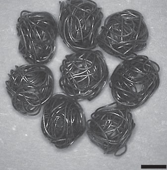
Entangled titanium fibre balls combined with nano strontium hydroxyapatite in repairing bone defects
- PMID: 24686383
- PMCID: PMC5586876
- DOI: 10.1159/000359951
Entangled titanium fibre balls combined with nano strontium hydroxyapatite in repairing bone defects
Abstract
Objective: To investigate the effect of entangled titanium fibre balls (ETFBs) combined with nano strontium hydroxyapatite (nano-Sr-HAP) on the repair of bone defects in vivo.
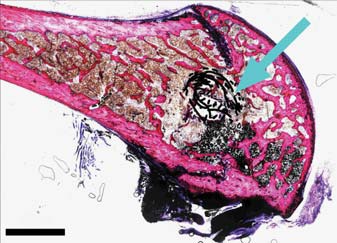
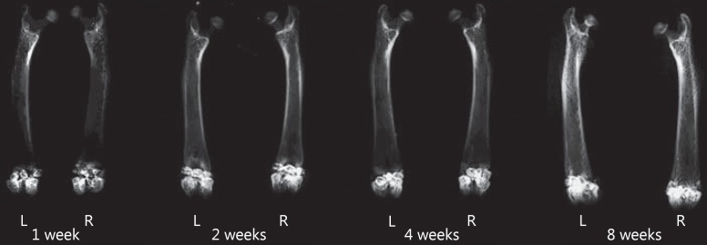
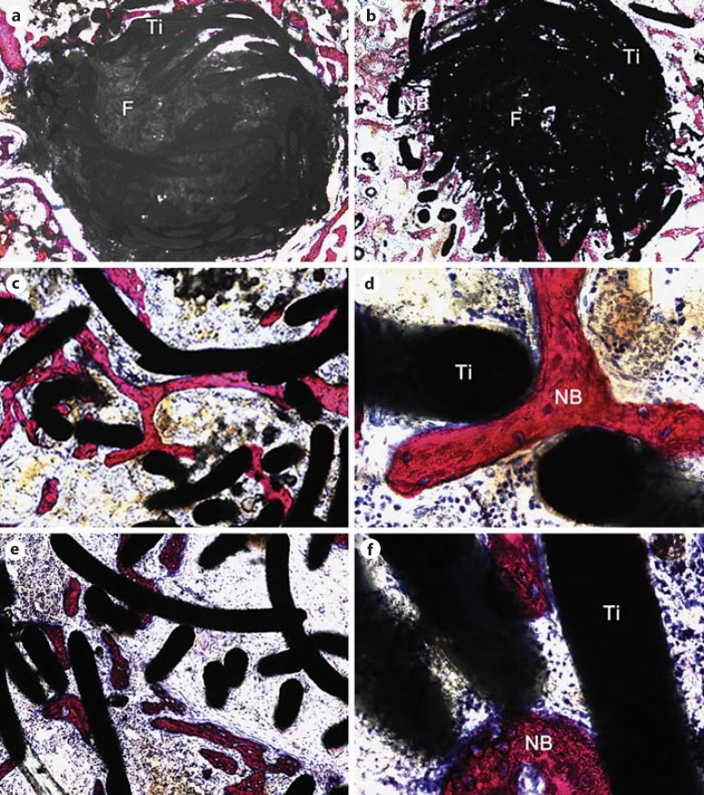
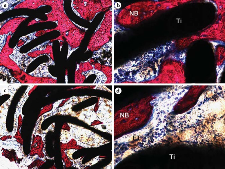
Materials and methods: Twenty-four 6-month-old, specific pathogen-free, male Sprague-Dawley rats were used. Drill defects were created in bilateral femoral condyles. ETFBs combined with nano-Sr-HAP were selected randomly from 72 samples and implanted into the femoral bone defects of left legs, which served as the experimental group, while ETFBs without nano-Sr-HAP were implanted into right legs for comparison. The bone defects on both sides were X-rayed. The anteroposterior positions and histological procedures and evaluations of each sample were performed at 1, 2, 4 and 8 weeks post-surgery.
Results: Histological results showed that the ETBs allowed new bone to grow within their structure. Additionally, an increase in new bone was seen on the nano-Sr-HAP side compared to the control side. The results of histomorphometric analysis confirmed that the new bone formation on the left side gradually increased with time. There was a statistical increase in new bone at 2, 4 and 8 weeks, and the differences between the two sides were statistically significant at weeks 4 and 8 (p < 0.05 for all comparisons).
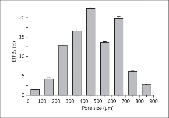
Conclusion: The results showed that ETFBs possess a unique 3-dimensional interconnective porous structure and have excellent biocompatibility, cell affinity and osteoconductivity, which makes them useful as scaffold materials for repairing bone defects. On the other hand, nano-Sr-HAP improved the bone defect-repairing capacity of the ETFBs, which showed osteoinductive properties.
Figures
References
-
- Takemoto M, Fujibayashi S, Neo M, et al. Mechanical properties and osteoconductivity of porous bioactive titanium. Biomaterials. 2005;26:6014–6023. - PubMed
-
- Kuboki Y, Takita H, Kobayashi D, et al. BMP-induced osteogenesis on the surface of hydroxyapatite with geometrically feasible and nonfeasible structures: topology of osteogenesis. J Biomed Mater Res. 1998;39:190–199. - PubMed
-
- Hutmacher DW. Scaffolds in tissue engineering bone and cartilage. Biomaterials. 2000;21:2529–2543. - PubMed
-
- Vehof JWM, van den Dolder J, de Ruijter JE, et al. Bone formation in Ca-P-coated and non-coated titanium fiber mesh. J Biomed Mater Res. 2003;64A:417–426. - PubMed
-
- Fassina L, Saino E, Visai L, et al. Electromagnetic enhancement of a culture of human SAOS-2 osteoblasts seeded onto titanium fiber-mesh scaffolds. J Biomed Mater Res A. 2008;87A:750–759. - PubMed
Publication types
MeSH terms
Substances
LinkOut - more resources
Full Text Sources
Other Literature Sources
Research Materials
