. 2014 Mar 31;9(3):e93497.
doi: 10.1371/journal.pone.0093497. eCollection 2014.

Solenopsis invicta virus 3: mapping of structural proteins, ribosomal frameshifting, and similarities to Acyrthosiphon pisum virus and Kelp fly virus

Affiliations

Solenopsis invicta virus 3: mapping of structural proteins, ribosomal frameshifting, and similarities to Acyrthosiphon pisum virus and Kelp fly virus

Steven M Valles et al. PLoS One. .

Abstract

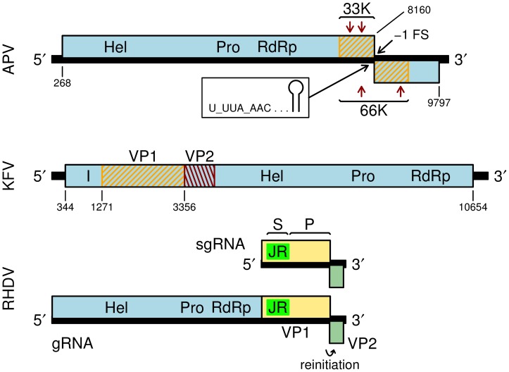
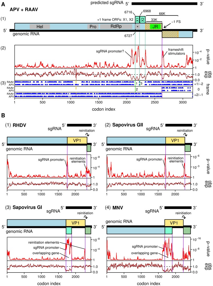
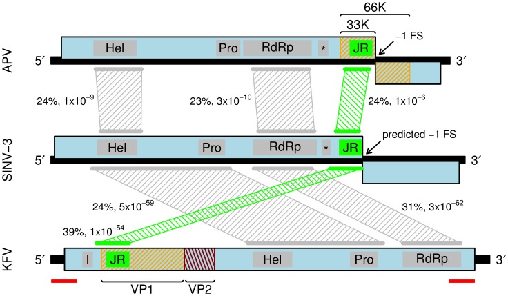
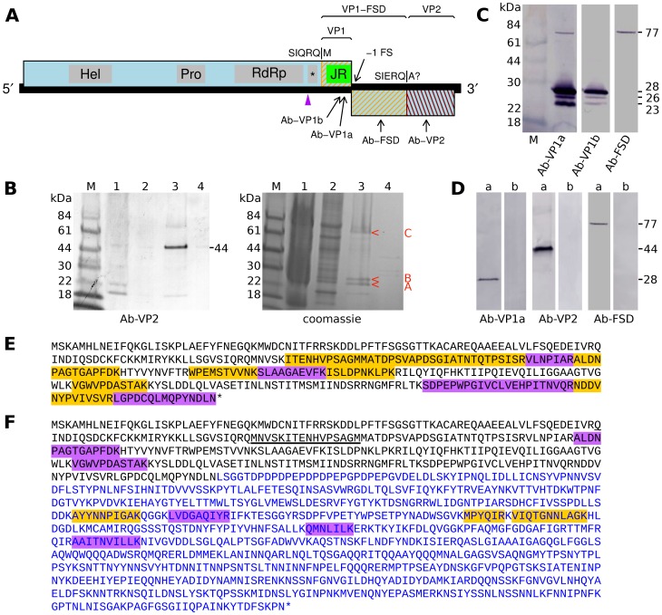
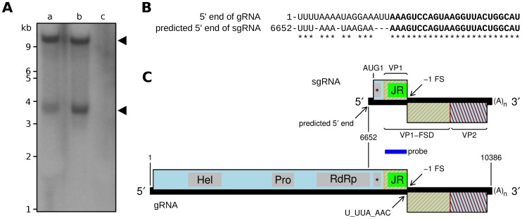
Solenopsis invicta virus 3 (SINV-3) is a positive-sense single-stranded RNA virus that infects the red imported fire ant, Solenopsis invicta. We show that the second open reading frame (ORF) of the dicistronic genome is expressed via a frameshifting mechanism and that the sequences encoding the structural proteins map to both ORF2 and the 3' end of ORF1, downstream of the sequence that encodes the RNA-dependent RNA polymerase. The genome organization and structural protein expression strategy resemble those of Acyrthosiphon pisum virus (APV), an aphid virus. The capsid protein that is encoded by the 3' end of ORF1 in SINV-3 and APV is predicted to have a jelly-roll fold similar to the capsid proteins of picornaviruses and caliciviruses. The capsid-extension protein that is produced by frameshifting, includes the jelly-roll fold domain encoded by ORF1 as its N-terminus, while the C-terminus encoded by the 5' half of ORF2 has no clear homology with other viral structural proteins. A third protein, encoded by the 3' half of ORF2, is associated with purified virions at sub-stoichiometric ratios. Although the structural proteins can be translated from the genomic RNA, we show that SINV-3 also produces a subgenomic RNA encoding the structural proteins. Circumstantial evidence suggests that APV may also produce such a subgenomic RNA. Both SINV-3 and APV are unclassified picorna-like viruses distantly related to members of the order Picornavirales and the family Caliciviridae. Within this grouping, features of the genome organization and capsid domain structure of SINV-3 and APV appear more similar to caliciviruses, perhaps suggesting the basis for a "Calicivirales" order.


Conflict of interest statement

Competing Interests: The authors have declared that no competing interests exist.

Figures

Figure 1
Figure 1. Genome maps for Acyrthosiphon pisum virus (APV), Kelp fly virus (KFV), and Rabbit hemorrhagic disease virus (RHDV).
RHDV is a calicivirus in the genus Lagovirus. Helicase (Hel), protease (Pro) and RNA-dependent RNA polymerase (RdRp) domains are indicated. A predicted inhibitor of apoptosis binding domain in KFV is indicated with an I . The APV structural proteins 33K and 66K (orange hatching; boundaries estimated based on protein sizes) are encoded near the 3' end of the genome; expression of 66K depends on a -1 ribosomal frameshift (-1 FS). Arrows indicate locations of 33K and 66K peptides detected via Edman degradation of cyanogen bromide cleavage products . The N-termini of the KFV structural proteins VP1 and VP2 have been mapped via Edman degradation; the C-terminal extent of VP2 is estimated from its gel mobility . The calicivirus structural proteins VP1 (tan) and VP2 (olive green) are expressed from a sgRNA. The N-terminal part of VP1 contains a picorna-like jelly-roll fold shell (S) domain ('JR'; bright green), while the C-terminal part comprises a protruding (P) domain.
Figure 2
Figure 2. Comparative genomic analysis of Acyrthosiphon pisum virus (APV) and Rosy apple aphid virus (RAAV).
A. (1) Map of the APV genome. The 33K structural protein contains a predicted picorna-like jelly-roll fold domain ('JR'; bright green). A predicted dsRNA binding domain is indicated with an asterisk. Potential overlapping genes X1 and X2 are indicated in turquiose. (2) Analysis of conservation at synonymous sites between APV and RAAV. The lower panel (brown) shows the ratio of the observed number of substitutions to the number expected under a null model of neutral evolution at synonymous sites, while the upper panel (red) shows the corresponding p-value. (3) Positions of stop codons (blue) in each of the three forward reading frames. Positions of alignment gaps are indicated in tan. The vertical green lines labeled '1', '2' and '3' indicate the positions and frames of the first three AUG codons on the predicted sgRNA. B. Genome maps and synonymous site conservation analyses for four calicivirus clades: (1) Rabbit hemorrhagic disease virus (RHDV; genus Lagovirus; 33 sequences), (2) group II sapoviruses (5 sequences), (3) group I sapoviruses (9 sequences), and (4) Murine norovirus (MNV; 58 sequences). VP1 and VP2 are indicated in tan and olive green, respectively; overlapping genes in group I sapoviruses and MNV are indicated in turquoise. All plots use a 25-codon sliding window, except MNV where a 15-codon sliding window is used. Note that p-values cannot be directly compared between plots as larger and more diverse alignments provide more statistical power (cf. the MNV and APV/RAAV plots).
Figure 3
Figure 3. Comparison of APV, SINV-3 and KFV genome organizations.
Mappings between homologous regions by simple blastp analysis are indicated with hatched trapezoids. Numbers indicate percentage amino acid identities and blastp e-values (e-values depend on subject database size and other variables so should be taken as indicative rather than absolute). SINV-3 is most similar to KFV despite KFV having a different genome organization. Predicted jelly-roll fold domains (JR) are indicated in bright green. Grey rectangles indicate the extent of domain sequences recognized by HHpred. Predicted dsRNA binding protein domains are indicated with an asterisk. Previously identified APV and KFV structural proteins are indicated with orange and red hatchings (see Fig. 1). Red bars indicate an unusual 638-nt almost-exact repeat sequence in the KFV published genome sequence (DQ112227.1).
Figure 4
Figure 4. Analysis of SINV-3 virion proteins.
A. Map of the SINV-3 genome indicating the locations of antibody 14-aa peptide antigens, the locations of structural proteins VP1, VP1-FSD and VP2 as revealed by Western analysis and mass spectrometry, and the N-terminus of VP1 as revealed by mass spectrometry. The cleavage site between VP1-FSD and VP2 has not been definitively localized but is close to the indicated site. B. Filtered extracts from SINV-3-infected worker ants were separated by centrifugation in a CsCl gradient (see Methods). Four bands were observed of which band 3 (sedimenting at 1.33 g/ml) was determined by qPCR to contain ∼100-fold more SINV-3 RNA than bands 1, 2 and 4 (Table 1). Analysis of the four fractions by SDS-PAGE (lanes 1–4) and Ab-VP2 Western analysis confirmed the presence of SINV-3 products mainly in fraction 3 (left, lane 3). Coomassie staining (right, lane 3) revealed specific products (i.e. relatively more intense in lane 3 than in other lanes) migrating at 77 kDa and in the 23–28 kDa range (red arrow heads). It is possible that other fractions (lanes 1, 2, 4) contain unrelated viruses present in the sample preparations. C. Western analysis of purified virion proteins with Ab-VP1a, Ab-VP1b and Ab-FSD, revealing VP1-related structural proteins. D. Western analysis of microsomal fractions prepared from infected worker ants. Samples from SINV-3-infected (a) and uninfected (b) ants were blotted and probed together and intervening lanes removed in silico for clarity. E. Tryptic peptides identified by mass spectrometry of band B (Fig. 4B, right, lane 3). The sequence region shown corresponds to the the C-terminal end of the ORF1 polyprotein, beginning from the purple arrow head in Fig. 4A. Peptides detected by mass spectrometry are highlighted; multiple highlight colours are used simply to distinguish adjacent peptides. F. Tryptic peptides identified by mass spectrometry of band C (Fig. 4B, right, lane 3). Black text represents amino acids encoded by the 3' end of ORF1; blue text represents amino acids encoded by ORF2. The N-terminal-most peptide detected in a chymotryptic digest of a gel slice containing the ∼77 kDa product is underlined; this was also the only N-terminally acetylated peptide detected.
Figure 5
Figure 5. Detection of a sgRNA species in SINV-3-infected ants.
A. Northern blot of (a) total RNA and (b) mRNA purified from SINV-3-infected Solenopsis invicta worker ants, and (c) total RNA purified from uninfected Solenopsis invicta worker ants. B. Alignment of the SINV-3 genomic and putative subgenomic 5' termini. A 24-nt perfect repeat sequence is indicated in bold. Genomic coordinates are indicated at left. C. Map of the SINV-3 genome indicating the sgRNA with its predicted 5' terminus and corresponding translation products. The region covered by the probe used in the Northern analysis is indicated by a dark blue bar.

Similar articles

Cited by

References

    1. van den Heuvel JF, Hummelen H, Verbeek M, Dullemans AM, van der Wilk F (1997) Characteristics of Acyrthosiphon pisum virus, a newly identified virus infecting the pea aphid. J Invertebr Pathol 70: 169–176. - PubMed
    1. van der Wilk F, Dullemans AM, Verbeek M, Van den Heuvel JF (1997) Nucleotide sequence and genomic organization of Acyrthosiphon pisum virus. Virology 238: 353–362. - PubMed
    1. Firth AE, Brierley I (2012) Non-canonical translation in RNA viruses. J Gen Virol 93: 1385–1409. - PMC - PubMed
    1. Scotti PD, Gibbs AJ, Wrigley NG (1976) Kelp fly virus. J Gen Virol 30: 1–9.
    1. Hartley CJ, Greenwood DR, Gilbert RJ, Masoumi A, Gordon KH, et al. (2005) Kelp fly virus: a novel group of insect picorna-like viruses as defined by genome sequence analysis and a distinctive virion structure. J Virol 79: 13385–13398. - PMC - PubMed

Publication types

LinkOut - more resources