Review
. 2014 Apr;13(4):290-314.
doi: 10.1038/nrd4228.

Advances in targeting cyclic nucleotide phosphodiesterases

Affiliations
Review

Advances in targeting cyclic nucleotide phosphodiesterases

Donald H Maurice et al. Nat Rev Drug Discov. 2014 Apr.

Abstract

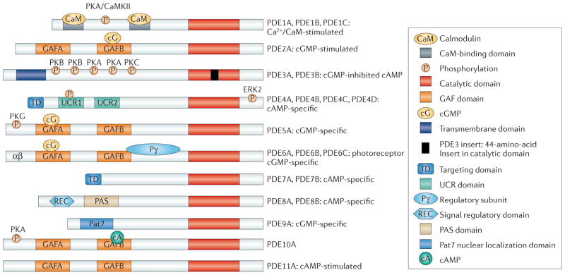
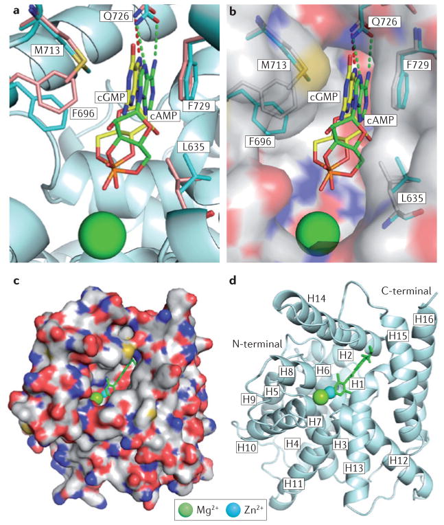
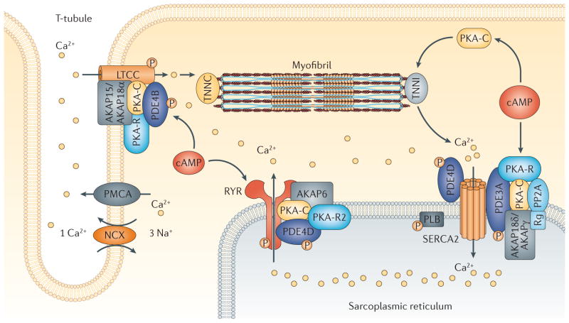
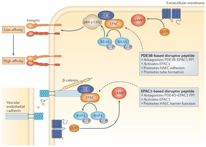
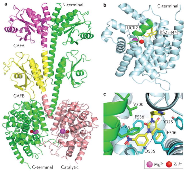
Cyclic nucleotide phosphodiesterases (PDEs) catalyse the hydrolysis of cyclic AMP and cyclic GMP, thereby regulating the intracellular concentrations of these cyclic nucleotides, their signalling pathways and, consequently, myriad biological responses in health and disease. Currently, a small number of PDE inhibitors are used clinically for treating the pathophysiological dysregulation of cyclic nucleotide signalling in several disorders, including erectile dysfunction, pulmonary hypertension, acute refractory cardiac failure, intermittent claudication and chronic obstructive pulmonary disease. However, pharmaceutical interest in PDEs has been reignited by the increasing understanding of the roles of individual PDEs in regulating the subcellular compartmentalization of specific cyclic nucleotide signalling pathways, by the structure-based design of novel specific inhibitors and by the development of more sophisticated strategies to target individual PDE variants.


Conflict of interest statement

Competing interests statement

The authors declare no competing interests.

Figures

Figure 1
Figure 1. Structure and domain organization of 11 mammalian PDE families
The conserved catalytic domain (shown in red) is located in the carboxy-terminal portion of the phosphodiesterases (PDEs). The catalytic domain of PDE3 contains a unique 44-amino-acid insert (shown in black). Many of the PDE families contain amino-terminal subdomains (such as GAF domains, transmembrane domains, targeting domains, upstream conserved regions (UCRs), PAS domains and REC domains) and N-terminal hydrophobic regions that are important in subcellular localization, in the incorporation of PDEs into compartmentalized signalosomes, in interactions with signalling molecules and molecular scaffolds, and in the regulation of PDE activity. GAF domains regulate the allosteric binding of cGMP (to PDE2, PDE5, PDE6 and PDE11), the allosteric binding of cAMP (to PDE10) and the regulation of catalytic activity (in PDE2, PDE5 and PDE6). Phosphorylation sites are labelled on the figure. CaMKII, calcium/calmodulin-dependent protein kinase II; ERK2, extracellular signal-regulated kinase 2; PKA, protein kinase A; Pat7, 7-residue nuclear localization signal.
Figure 2
Figure 2. Binding of substrates and inhibitors to the active site of PDEs
a | Interaction of the substrates cAMP (green sticks) and cGMP (yellow sticks) with the D674A mutant of phosphodiesterase 10A2 (PDE10A2). Residues from the structures of PDE10A2–cAMP and PDE10A2–cGMP are shown in cyan and pale orange, respectively. Binding to Zn2+ was lost owing to the D674A mutation. Substrates of cAMP and cGMP have the same syn-conformation, but opposite orientations. b | Surface presentation of the binding of cAMP and cGMP to the D674A mutant of PDE10A2. c | Surface presentation of PDE4D2 binding to roflumilast (green stick). d | Ribbon diagram of PDE4D2 bound to roflumilast. ‘H’ represents the helical loops in the catalytic core. See REFS ,,, for more information.
Figure 3
Figure 3. PDE-containing signalosomes couple cAMP–PKA signalling to myocardial contractility
Phosphodiesterase 4B (PDE4B), as a component of an A-kinase anchor protein 15/18 (AKAP15/AKAP18α)-based signalosome in the cardiac L-type calcium channel (LTCC) complex, regulates the calcium current and protects against ventricular arrythmias in mice. AKAP6 (also known as mAKAP) serves as a scaffold for a PDE4D3-containing signalosome that is involved in regulating the release of calcium from the sarcoplasmic reticulum via the ryanodine receptor channel. PDE3A, as a component of a sarcoplasmic/endoplasmic reticulum calcium ATPase (SERCA) AKAP18δ-containing signalosome, is an important regulator of the effects of cAMP on calcium uptake into the sarcoplasmic reticulum. NCX, sodium/calcium exchanger; PKA, protein kinase A; PKA-C, PKA catalytic domain; PKA-R, PKA regulatory domain; PLB, phospholamban; PMCA, plasma membrane calcium-transporting ATPase; PP2A, protein phosphatase 2A; Rg, PP2A regulatory subunit; TNNC, troponin C; TNNI, troponin I.
Figure 4
Figure 4. Human arterial endothelial cells construct two distinct PDE- and EPAC-based signalosomes to regulate, spatially and functionally, integrin- and vascular endothelial cadherin-based adhesions, respectively
As shown in the top panel, a direct interaction between phosphodiesterase 3B (PDE3B) and exchange factor directly activated by cAMP (EPAC) promotes the formation of a plasma membrane signalosome that controls cAMP-mediated activation of RAS-related protein (RRAS) and its subsequent activation of the p84-regulatory domain-associated p110γ subunit of phosphoinositide 3-kinase γ and, ultimately, integrin-based adhesions with the extracellular matrix. A PDE3B-based, EPAC1-displacing peptide antagonizes PDE3B–EPAC1 protein–protein interactions (PPIs), allows localized increases in cAMP and promotes signalling through this signalosome. As shown in the bottom panel, a direct interaction between PDE4D and EPAC1 promotes the integration of these proteins into a vascular endothelial cadherin-based signalosome that allows cAMP — acting through EPAC1-based activation of small GTPase RAS-related protein 1 (RAP1) — to regulate endothelial cell permeability. An EPAC1-based, PDE4D-displacing peptide antagonizes PDE4D–EPAC1 PPIs, interferes with the integration of these proteins into the signalosome and increases endothelial cell permeability. HAEC, human arterial endothelial cell; PIP3, phosphatidylinositol-(3,4,5)-trisphosphate; PKB, protein kinase B.
Figure 5
Figure 5. Allosteric modulation of PDE2 and PDE4
a | Ribbon diagram of near-full-length unliganded phosphodiesterase 2A (PDE2A). The entire molecule A of the PDE2A dimer is shown in green, whereas GAF domain A (GAFA) is shown in magenta, GAFB in yellow and the catalytic domain of molecule B in pale pink,. b | Ribbon diagram of the PDE4 catalytic domain (shown in pale cyan) with an upstream conserved region 2 (UCR2) fragment (shown in green). The PDE4-selective inhibitor RS25344 (shown in yellow) binds to the active site of PDE4 and also interacts with the UCR2 fragment. c | Detailed view of the interaction of RS25344 (shown in yellow) with PDE4D3 residues (shown in cyan and green; Protein Data Bank ID: 3G4G). Similar to other PDE4 inhibitors, the allosteric modulator RS25344 forms a conserved hydrogen bond with the invariant Gln535 and stacks against Phe538 in the catalytic pocket. However, RS25344 makes unique hydrophobic contacts with Phe196 and Val200 in the UCR2 helix, which ‘caps’ and interacts with the active site,.

Similar articles

Cited by

References

    1. Conti M, Beavo J. Biochemistry and physiology of cyclic nucleotide phosphodiesterases: essential components in cyclic nucleotide signaling. Annu Rev Biochem. 2007;76:481–511. - PubMed
    1. Francis S, Blount M, Corbin J. Mammalian cyclic nucleotide phosphodiesterases: molecular mechanisms and physiological functions. Physiol Rev. 2011;91:651–690. This is an excellent, comprehensive and very timely review of PDEs. - PubMed
    1. Keravis T, Lugnier C. Cyclic nucleotide phosphodiesterase (PDE) isozymes as targets of the intracellular signalling network: benefits of PDE inhibitors in various diseases and perspectives for future therapeutic developments. Br J Pharmacol. 2012;165:1288–1305. This is an excellent and concise review of PDEs. - PMC - PubMed
    1. Derbyshire E, Marletta M. Structure and regulation of soluble guanylate cyclase. Annu Rev Biochem. 2012;81:533–559. - PubMed
    1. Potter LR. Regulation and therapeutic targeting of peptide-activated receptor guanylyl cyclases. Pharmacol Ther. 2011;130:71–82. - PMC - PubMed

Publication types