. 2014 May 9;114(10):1576-84.
doi: 10.1161/CIRCRESAHA.114.302313. Epub 2014 Mar 31.

Disruption of mammalian target of rapamycin complex 1 in macrophages decreases chemokine gene expression and atherosclerosis

Affiliations

Disruption of mammalian target of rapamycin complex 1 in macrophages decreases chemokine gene expression and atherosclerosis

Ding Ai et al. Circ Res. .

Abstract

Rationale: The mammalian target of rapamycin complex 1 inhibitor, rapamycin, has been shown to decrease atherosclerosis, even while increasing plasma low-density lipoprotein levels. This suggests an antiatherogenic effect possibly mediated by the modulation of inflammatory responses in atherosclerotic plaques.

Objective: Our aim was to assess the role of macrophage mammalian target of rapamycin complex 1 in atherogenesis.

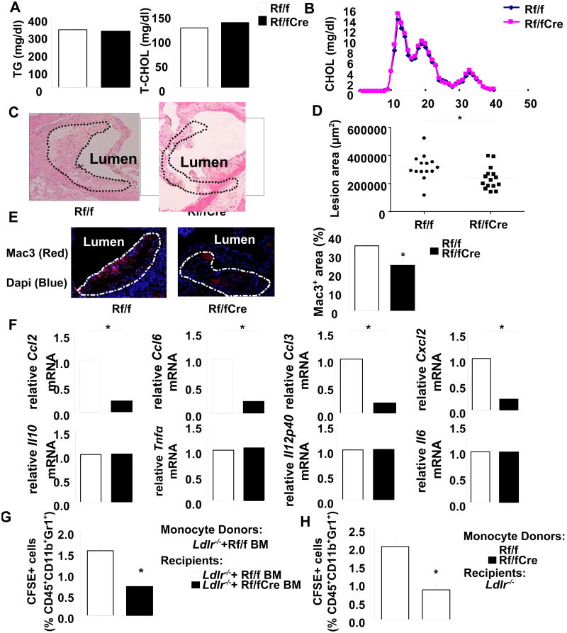
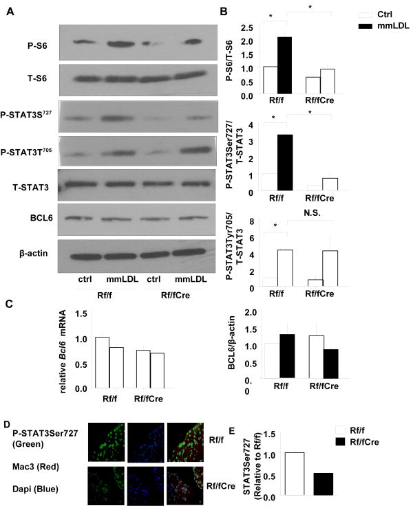
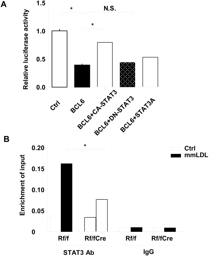
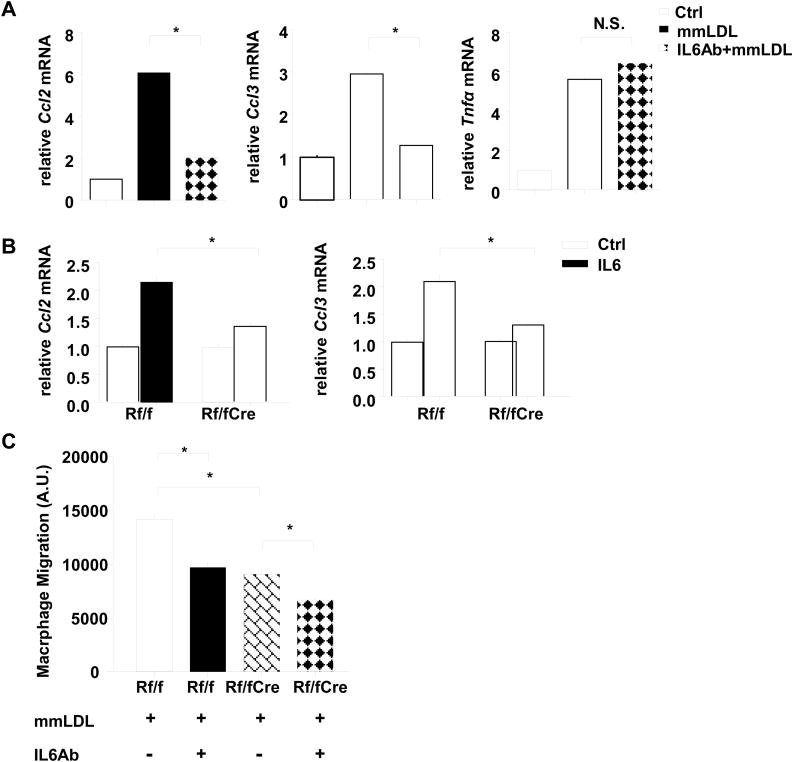
Methods and results: We transplanted bone marrow from mice in which a key mammalian target of rapamycin complex 1 adaptor, regulatory-associated protein of mTOR, was deleted in macrophages by Cre/loxP recombination (Mac-Rap(KO) mice) into Ldlr(-/-) mice and then fed them the Western-type diet. Atherosclerotic lesions from Mac-Rap(KO) mice showed decreased infiltration of macrophages, lesion size, and chemokine gene expression compared with control mice. Treatment of macrophages with minimally modified low-density lipoprotein resulted in increased levels of chemokine mRNAs and signal transducer and activator of transcription (STAT) 3 phosphorylation; these effects were reduced in Mac-Rap(KO) macrophages. Although wild-type and Mac-Rap(KO) macrophages showed similar STAT3 phosphorylation on Tyr705, Mac-Rap(KO) macrophages showed decreased STAT3Ser727 phosphorylation in response to minimally modified low-density lipoprotein treatment and decreased Ccl2 promoter binding of STAT3.

Conclusions: The results demonstrate cross-talk between nutritionally induced mammalian target of rapamycin complex 1 signaling and minimally modified low-density lipoprotein-mediated inflammatory signaling via combinatorial phosphorylation of STAT3 in macrophages, leading to increased STAT3 activity on the chemokine (C-C motif) ligand 2 (monocyte chemoattractant protein 1) promoter with proatherogenic consequences.

Keywords: atherosclerosis; macrophages; mammalian target of rapamycin complex 1.


Figures

Figure 1
Figure 1. Plasma lipid levels and atherosclerotic lesions in the aortic root of Ldlr-/- mice transplanted with Raptorflox/flox or Mac-RapKO BM
Mice were fed WTD for 10 weeks. A. Plasma triglycerides and total cholesterol levels were measured in Ldlr-/- mice with Raptorflox/flox (Rf/f) or Mac-RapKO (Rf/fCre) BM transplantation. N=5-8. B. Cholesterol lipoprotein distribution as determined by fast performance liquid chromatography on pooled plasma samples. N=5-8 per pool. C. H&E staining of representative aortic root sections. Atherosclerotic lesions are demarcated by the dashed lines. N=14-15. D. Quantification of lesions by morphometric analysis. E. Sections were stained with a Mac3 antibody for the presence of macrophages (left), and values represent the percentages of Mac3+ area/total lesion area (right). N=5. F. mRNA levels of the indicated targets were assayed in LCM-captured RNA obtained from atherosclerotic lesions. The data were normalized to the expression level of β-actin mRNA. N=5-7. *P<0.05. (G) Classical monocytes sorted from Ldlr-/- mice transplanted with Rapflox/flox BM were labeled with CFSE, and injected intravenously into Ldlr-/- mice that had been transplanted with BM from either Rapflox/flox or Mac-RapKO mice ; (H) monocytes from Rapflox/flox or Mac-RapKO mice were transplanted into Ldlr-/- mice. CFSE labeled cells (as a percentage of CD45+CD11b+Gr1+ cells) in the aorta were quantified by flow cytometry. N=5. *P < 0.05.
Figure 2
Figure 2. mmLDL mediated inflammatory gene expression in bone marrow derived macrophages (BMDMs) from LysmCre, Raptorflox/flox and Mac-RapKO mice
A. BMDMs from LysmCre, Raptorflox/flox (Rf/f) and Mac-RapKO mice (Rf/fCre) were treated with mmLDL (50ug/ml) for 2 hours. Expression levels of Tnfα, Il6, Il10 and Il12p40 were measured by quantitative -PCR and normalized to expression levels observed without treatment (Ctrl). B. Ccl2, Ccl3, Ccl6, Ccl7 and Cxcl2 gene expression in BMDMs of LysmCre, Rf/f and Rf/fCre mice after 2 hours of mmLDL (50 μg/ml) treatment. N=3, *P<0.05 C. BMDMs were treated with mmLDL for 2 hours, washed with PBS and then incubated with fresh media for another 18 hours. CCL2 in the media was measured by ELISA and values were normalized to total cellular protein. N=3, *P<0.05, N.S., not significant
Figure 3
Figure 3. Regulation of STAT3 phosphorylation by Raptor deficiency in BMDMs
A. Western blot analysis of S6 and STAT3 phosphorylation and BCL6 expression. β-Actin was used as internal control. P-, phosphorylated; T-, total B. Quantification of protein levels N=3, *P < 0.05. Rf/f vs. Rf/f+mmLDL, Rf/f+mmLDL vs. Rf/fCre+mmLDL, one-way ANOVA, Bonferroni post-test. N.S., not significant C. Measurement of macrophage Bcl6 mRNA by qPCR. N=3 D. Sections of the aortic root were double-stained with Mac3 and phospho-STAT3Ser727 antibodies. E. Relative fluorescence intensity for phospho-STAT3Ser727 expression (green) in the macrophage-dense areas (red) of the lesions is shown. Quantification of phospho-STAT3Ser727 in lesion macrophage area normalized to the value of Rf/f. N=5. *P < 0.05.
Figure 4
Figure 4. Mechanism of CCl2 regulation by Raptor
A. Ccl2 promoter inserted into a reporter system was cotransfected with CA-STAT3, DN-STAT3 or STAT3A plasmids in the presence or absence of Bcl6 plasmid in 293 cell lines. An empty vectors were used as normalization control. N=3, *P < 0.05. NS, not significant B. ChIP-qPCR analysis of STAT3 binding on the Ccl2 genomic locus in BMDMs from Rf/f or Rf/fCre mice with or without mmLDL treatment using STAT3 antibody or rabbit IgG. The fold-enrichment of the Ccl2 locus was determined by qPCR and calculated as percentage of input. Data are from three independent experiments. N=3, *P < 0.05.
Figure 5
Figure 5. Chemokine expression and migratory response induced by mmLDL was blocked by IL6 neutralizing antibody in BMDMs from wild type mice
A. Expression levels of chemokines and Tnfα were measured by quantitative RT-PCR in wild type BMDMs 2 hours after mmLDL (50 μg/ml) treatment with or without a 1 hour rat IgG against IL-6 or isotype control (0.5 μg/ml) pretreatment and normalized to Ctrl. N=3 B. BMDMs from Rf/f and Rf/fCre mice were treated with IL6 (20 ng/ml) for 2 hours. Expression levels of chemokines were measured by quantitative PCR and normalized to expression levels observed without treatment. N=3 C. Macrophage migration assay was performed in which BMDMs from Rf/f and Rf/fCre mice were added to bottom wells and pre-treated with mmLDL (50 μg/ml) and rat IgG against IL-6 or isotype control for 24 hours. The migrated macrophages were quantified by fluorescence spectroscopy. N=3, *P < 0.05. one-way ANOVA, Bonferroni post-test. N.S., not significant.

References

    1. Kapahi P, Chen D, Rogers AN, Katewa SD, Li PW, Thomas EL, Kockel L. With tor, less is more: A key role for the conserved nutrient-sensing tor pathway in aging. Cell Metab. 2010;11:453–465. - PMC - PubMed
    1. Inoki K, Li Y, Xu T, Guan KL. Rheb gtpase is a direct target of tsc2 gap activity and regulates mtor signaling. Genes Dev. 2003;17:1829–1834. - PMC - PubMed
    1. Hara K, Maruki Y, Long X, Yoshino K, Oshiro N, Hidayat S, Tokunaga C, Avruch J, Yonezawa K. Raptor, a binding partner of target of rapamycin (tor), mediates tor action. Cell. 2002;110:177–189. - PubMed
    1. Kim DH, Sarbassov DD, Ali SM, King JE, Latek RR, Erdjument-Bromage H, Tempst P, Sabatini DM. Mtor interacts with raptor to form a nutrient-sensitive complex that signals to the cell growth machinery. Cell. 2002;110:163–175. - PubMed
    1. Guertin DA, Stevens DM, Thoreen CC, Burds AA, Kalaany NY, Moffat J, Brown M, Fitzgerald KJ, Sabatini DM. Ablation in mice of the mtorc components raptor, rictor, or mlst8 reveals that mtorc2 is required for signaling to akt-foxo and pkcalpha, but not s6k1. Dev Cell. 2006;11:859–871. - PubMed

Publication types

MeSH terms