. 2014 Mar 31;204(7):1219-36.
doi: 10.1083/jcb.201307057.

Functionally distinct PI 3-kinase pathways regulate myelination in the peripheral nervous system

Affiliations

Functionally distinct PI 3-kinase pathways regulate myelination in the peripheral nervous system

Bradley A Heller et al. J Cell Biol. .

Abstract

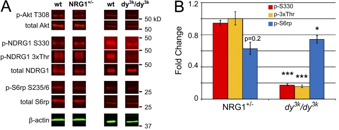
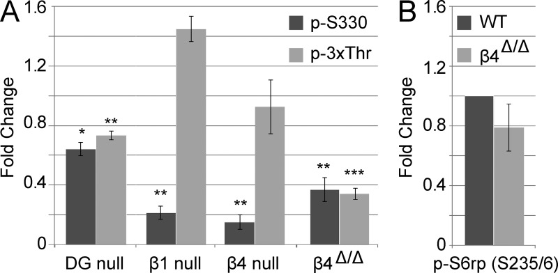
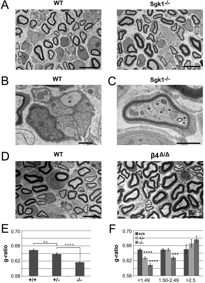
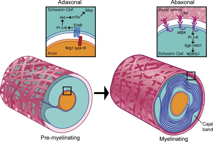
The PI 3-kinase (PI 3-K) signaling pathway is essential for Schwann cell myelination. Here we have characterized PI 3-K effectors activated during myelination by probing myelinating cultures and developing nerves with an antibody that recognizes phosphorylated substrates for this pathway. We identified a discrete number of phospho-proteins including the S6 ribosomal protein (S6rp), which is down-regulated at the onset of myelination, and N-myc downstream-regulated gene-1 (NDRG1), which is up-regulated strikingly with myelination. We show that type III Neuregulin1 on the axon is the primary activator of S6rp, an effector of mTORC1. In contrast, laminin-2 in the extracellular matrix (ECM), signaling through the α6β4 integrin and Sgk1 (serum and glucocorticoid-induced kinase 1), drives phosphorylation of NDRG1 in the Cajal bands of the abaxonal compartment. Unexpectedly, mice deficient in α6β4 integrin signaling or Sgk1 exhibit hypermyelination during development. These results identify functionally and spatially distinct PI 3-K pathways: an early, pro-myelinating pathway driven by axonal Neuregulin1 and a later-acting, laminin-integrin-dependent pathway that negatively regulates myelination.


Figures

Figure 1.
Figure 1.
Phospho-substrates are up-regulated with myelination. (A) Myelinating co-cultures of rat sensory neurons and Schwann cells stained for MBP (red), p-Sub (green), and laminin (white); boxed areas are shown at higher magnification below at 1.5×. Myelinating Schwann cells (inset, arrow) are selectively stained with the phospho-substrate antibody (p-Sub). Some Schwann cells that have elongated but are not yet expressing MBP at significant levels are stained with the p-Sub antibody (arrowheads). Bar, 75 µm. (B) Staining of myelinating co-cultures for p-Sub (green), POU3F1 (red), and MBP (blue) is shown. A subset of premyelinating POU3F1-expressing Schwann cells coexpress phospho-substrates (arrow). All MBP-expressing Schwann cells, which are POU3F1-negative, coexpress phospho-substrates (arrowheads). Bar, 10 µm. (C) Myelinating co-cultures stained with p-Sub (green), Mupp1 (red), and MBP (blue) indicate that phospho-substrates are enriched in the SLI. Bar, 5 µm. (D) Myelinating co-cultures stained with p-Sub (green), Caspr1 (red), and MBP (blue) indicate that phospho-substrates are sometimes enriched in the distal glial paranode (arrow), extending into the juxtaparanode (arrowhead). Bar, 5 µm.
Figure 2.
Figure 2.
Phospho-substrates are differentially expressed during myelination. (A) Time course of the expression of phospho-substrates in myelinating co-cultures. Western blot analysis of lysates prepared from co-cultures beginning on day 0, when myelinating media was added, through day 21. Six major bands were detected that either increased or decreased as myelination proceeded. The p-Sub reactive bands are in red, β-actin in green; P0 myelin protein expression is also shown. (B) Co-cultures of Schwann cells and sensory neurons were maintained for 35 d under nonmyelinating (NM) or myelinating (Myel.) conditions. The increase in p45 and decrease in p30 observed in the time course shown in A only occur under myelinating conditions.
Figure 3.
Figure 3.
S6rp and NDRG1 correspond to the 30- and 45-kD phospho-substrate bands. (A) Western blots of wild-type and stretcher sciatic nerve lysates were probed with anti-NDRG1 and p-Sub antibodies; β-actin serves as a loading control. NDRG1 and the p45-kD p-Sub doublet are not detected in the stretcher nerve lysates. (B) Western blots of lysates from nonmyelinating co-cultures were treated with 50 nM rapamycin, 20 µM LY294002, or 40 µM PF-4708671 for 18 h and then probed with p-Sub, p-S6rp, and total S6rp antibodies. p-S6rp and p30 are not detectable in co-cultures treated with rapamycin, LY294002, or PF-4708671. (C) Expression of phospho- and total Akt, NDRG1, and S6rp in developing rat sciatic nerves. Lysates of postnatal sciatic nerves were probed on Western blots with the indicated antibodies. Both total and phosphorylated NDRG1 are up-regulated in parallel with compact myelin proteins P0 and MBP. S6rp and Akt are maximally expressed and phosphorylated early, then down-regulated as myelination proceeds.
Figure 4.
Figure 4.
Localization of p-S6rp, p-NDRG1, and p-Akt in myelinating co-cultures. (A) Co-cultures maintained in myelinating media for 5 or 14 d were stained for p-Akt (green), Caspr (red), and MBP (blue). In 5-d co-cultures (top row), Schwann cells just before myelinating express p-Akt (S473) along their length; Caspr staining of axons is diffuse and MBP is not yet expressed. In 14-d co-cultures (bottom two rows), p-Akt staining varied depending on the maturity of the myelin segments. In a thinly myelinated segment with a single paranode (bracket), p-Akt was prominent along the Schwann cells, in more mature segments with two hemi-paranodes (arrows) or surrounding a mature node (arrowheads) staining was concentrated in the paranodal and juxtaparanodal regions. Addition of the LY inhibitor before fixation abolished p-Akt staining. Bar, 20 µm. (B) Myelinating co-cultures were stained for p-S6rp (green), POU3F1 (red), and MBP (blue). p-S6rp was maximally expressed in the cytoplasm and along the processes of a premyelinating Schwann cell expressing POU3F1 (arrows) and at lower levels in myelinated Schwann cells (one is marked by an asterisk). Bar, 20 µm. Inset (far right panel) is at 2×. (C) Myelinating co-cultures stained for phospho- (3xThr, S330) and total NDRG1 (all green), MBP (blue), and Caspr (red). Myelinating Schwann cells express phospho- and total NDRG1 (green) along the outside of the myelin sheath and in some paranodes (insets, 3× magnification). Bar, 20 µm.
Figure 5.
Figure 5.
NDRG1 is enriched in the Cajal bands of the abaxonal compartment. (A) A teased adult rat sciatic nerve fiber stained for total NDRG1 (green) and Necl-4 (red) is shown. Total NDRG1 is present in the abaxon (green arrow), adaxon (yellow arrow), and throughout the length of the SLI (white brackets); Necl-4 is present in the adaxonal membrane and the incisures. Inset is rotated and shown at 3×. Bar, 20 µm. (B) Teased adult rat sciatic nerve fiber stained for phospho-NDRG1 (green) and Necl-4 (red) is shown. p-NDRG1 is expressed in the abaxon (green arrow) but not the adaxon (red arrow) and only the outer portion of the SLI (white brackets). Necl-4 is expressed throughout the entire incisures. A Cajal band is present in the center of the fiber (white asterisks). Inset is rotated and shown at 3×. Bar, 20 µm. (C and D) Teased P45 mouse sciatic nerve fibers stained for (C) total or (D) p-NDRG1 (green) and α-dystroglycan (red); SLI are labeled with white arrowheads. Both total and p-NDRG1 are enriched in the Cajal bands and excluded from the α-DG stained appositions. Bar, 20 µm.
Figure 6.
Figure 6.
Neuregulin1 and laminin-2 differentially regulate S6rp and NDRG1 phosphorylation. (A) Lysates of P10 nerve from Neuregulin1 type III haploinsufficient or α2-laminin–null (dy3k/dy3k) mice were blotted for total and phosphorylated Akt, S6rp, and NDRG1. Whereas p-Akt is reduced in both nerves, phospho-NDRG1 is markedly reduced only in the α2-laminin–null mice. (B) Quantification of the phosphoprotein levels in neuregulin and laminin mutant nerves are shown normalized to total protein in each case. Mean ± SEM; *, P < 0.05; ***, P < 0.001 by two-tailed Student’s t test; n ≥ 3.
Figure 7.
Figure 7.
β4 integrin is a key laminin receptor upstream of NDRG1 phosphorylation. (A) Lysates from P10 nerves deficient in various laminin receptors were probed for alterations in NDRG1 phosphorylation at the p-S330 and p-3xThr sites. p-NDRG1 levels are normalized to total NDRG1 for each quantification. Conditional ablation of the β1 (n = 3) and β4 (n = 4) integrin subunit causes a defect on S330 phosphorylation but not p-3xThr. The signaling-defective β4 mutant (β4Δ/Δ; n = 7) exhibits a marked reduction of phosphorylation at both the S330 and 3xThr sites. The DG-null has a small but significant defect at both S330 and 3xThr (n = 5). Mean ± SEM; *, P ≤ 0.05; **, P < 0.01; ***, P < 0.001 by two-tailed t test. (B) P10 β4-signaling mutant nerves have normal levels of S6rp phosphorylation. Quantifications were normalized to total S6rp protein levels. Mean ± SEM; n = 4; P > 0.05 by two-tailed t test.
Figure 8.
Figure 8.
Sgk isoforms phosphorylate NDRG1. (A) RT-PCR of first-strand cDNA obtained from pure sensory neurons, Schwann cells, and kidney (control) using Sgk isoform-specific primers demonstrates Sgk1 and Sgk3 are expressed by Schwann cells. (B) Quantification of Western blots analyzing phosphorylation at the p-S330 and 3xThr sites of NDRG1 in P10 and adult (∼P60) sciatic nerves from Sgk1- or Sgk3-specific knockouts. Sgk1 loss of function reduces NDRG1 phosphorylation at the 3xThr site at P10 and at both sites in the adult. Sgk3 loss of function slightly increases p-S330 and decreases 3xThr in the adult. p-NDRG1 was normalized to total NDRG1, which was similar across phenotypes, for the quantification. Mean fold change relative to wild type ± SEM, n ≥ 4. *, P < 0.05; ***, P < 0.001 by two-tailed Student’s t test.
Figure 9.
Figure 9.
Sgk1-null mice are hypermyelinated during development. (A) Representative electron microscopic fields of P10 nerves from wild-type and Sgk1−/− mice are shown, demonstrating modest hypermyelination of some fibers in the null nerve. Bars, 5 µm. (B) At P10, large-caliber axons (asterisk) are still occasionally ensheathed in the Remak bundles of wild-type but not Sgk1 knockout nerves (not depicted). Bar, 1 µm. (C) An example of multiple small axons (asterisks) communally myelinated by a single Schwann cell from the Sgk1-null mice is shown. Bar, 1 µm. (D) Representative electron microscopic fields of P10 nerves from wild-type and β4-signaling mutant mice are shown, demonstrating modest hypermyelination of some fibers in the mutant nerve. Bars, 5 µm. (E) Quantification of the g-ratios in P10 nerves from wild-type (884 axons scored), Sgk1+/− (942 axons scored), and Sgk1−/− (796 axons scored) mice. Loss of Sgk1 results in a modest hypermyelination that is dose dependent. Mean g-ratios are shown ± SEM; **, P < 0.01; ****, P < 0.0001 by ANOVA. (F) Quantification of the g-ratios in P10 nerves from wild type, Sgk1+/−, and Sgk1−/− binned by axon caliber. Mean g-ratios are shown ± SEM; ***, P < 0.001; ****, P < 0.0001 by ANOVA and Tukey’s multiple comparisons test.
Figure 10.
Figure 10.
Schematic of PI 3-K pathways activated during myelination. Schematic of the proposed transition in PI 3-K during myelination. In premyelinating Schwann cells (left), Neuregulin1 drives myelination by activating TORC1 through erbB-dependent PI 3-K/Akt activation. In myelinating Schwann cells (right), pro-myelinating signals are opposed by laminin signaling via α6β4 integrin and Sgk1. This signaling is localized to the Cajal bands of the abaxon; among the targets of Sgk1 phosphorylation is NDRG1, a protein required for long-term myelin maintenance.

References

    1. Alessi D.R., Caudwell F.B., Andjelkovic M., Hemmings B.A., Cohen P. 1996. Molecular basis for the substrate specificity of protein kinase B; comparison with MAPKAP kinase-1 and p70 S6 kinase. FEBS Lett. 399:333–338 10.1016/S0014-5793(96)01370-1 - DOI - PubMed
    1. Alessi D.R., Deak M., Casamayor A., Caudwell F.B., Morrice N., Norman D.G., Gaffney P., Reese C.B., MacDougall C.N., Harbison D., et al. 1997a. 3-Phosphoinositide-dependent protein kinase-1 (PDK1): structural and functional homology with the Drosophila DSTPK61 kinase. Curr. Biol. 7:776–789 10.1016/S0960-9822(06)00336-8 - DOI - PubMed
    1. Alessi D.R., James S.R., Downes C.P., Holmes A.B., Gaffney P.R., Reese C.B., Cohen P. 1997b. Characterization of a 3-phosphoinositide-dependent protein kinase which phosphorylates and activates protein kinase Balpha. Curr. Biol. 7:261–269 10.1016/S0960-9822(06)00122-9 - DOI - PubMed
    1. Atanasoski S., Scherer S.S., Sirkowski E., Leone D., Garratt A.N., Birchmeier C., Suter U. 2006. ErbB2 signaling in Schwann cells is mostly dispensable for maintenance of myelinated peripheral nerves and proliferation of adult Schwann cells after injury. J. Neurosci. 26:2124–2131 10.1523/JNEUROSCI.4594-05.2006 - DOI - PMC - PubMed
    1. Bae D.H., Jansson P.J., Huang M.L., Kovacevic Z., Kalinowski D., Lee C.S., Sahni S., Richardson D.R. 2013. The role of NDRG1 in the pathology and potential treatment of human cancers. J. Clin. Pathol. 66:911–917 10.1136/jclinpath-2013-201692 - DOI - PubMed

Publication types

MeSH terms