Review
. 2014 Aug;71(16):3139-50.
doi: 10.1007/s00018-014-1610-7. Epub 2014 Apr 1.

Transport and diffusion of Tau protein in neurons

Affiliations
Review

Transport and diffusion of Tau protein in neurons

Tim Scholz et al. Cell Mol Life Sci. 2014 Aug.

Abstract

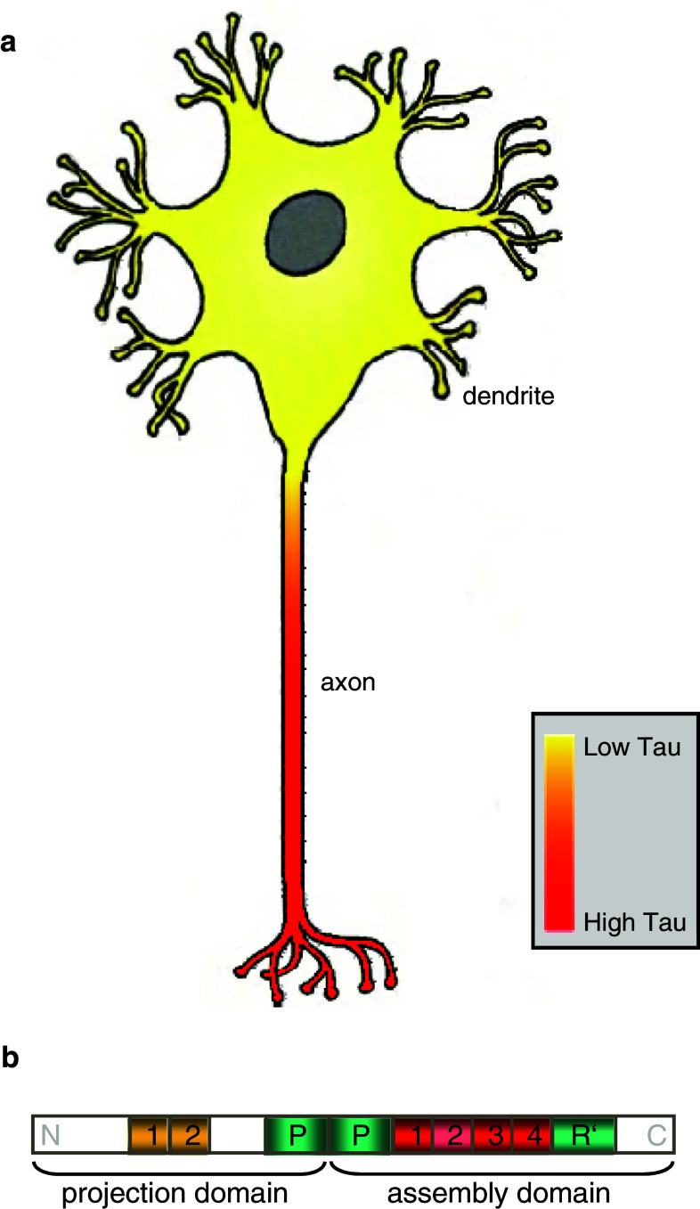
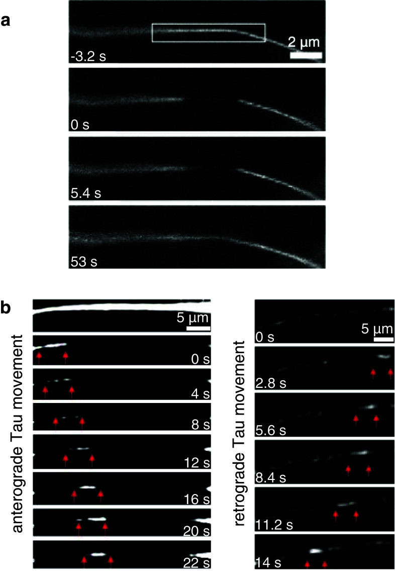
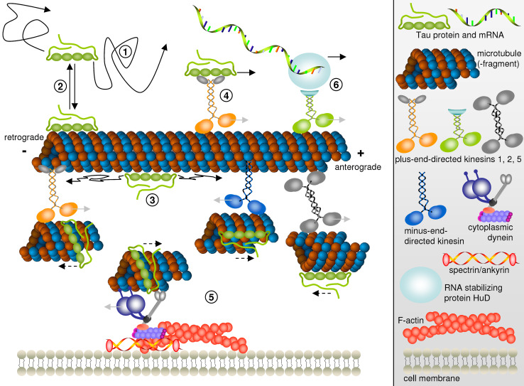
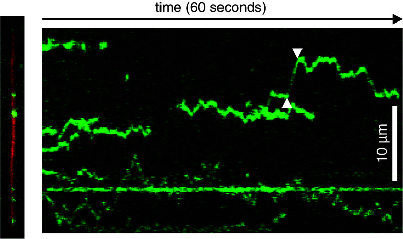
In highly polarized and elongated cells such as neurons, Tau protein must enter and move down the axon to fulfill its biological task of stabilizing axonal microtubules. Therefore, cellular systems for distributing Tau molecules are needed. This review discusses different mechanisms that have been proposed to contribute to the dispersion of Tau molecules in neurons. They include (1) directed transport along microtubules as cargo of tubulin complexes and/or motor proteins, (2) diffusion, either through the cytosolic space or along microtubules, and (3) mRNA-based mechanisms such as transport of Tau mRNA into axons and local translation. Diffusion along the microtubule lattice or through the cytosol appear to be the major mechanisms for axonal distribution of Tau protein in the short-to-intermediate range over distances of up to a millimetre. The high diffusion coefficients ensure that Tau can distribute evenly throughout the axonal volume as well as along microtubules. Motor protein-dependent transport of Tau dominates over longer distances and time scales. At low near-physiological levels, Tau is co-transported along with short microtubules from cell bodies into axons by cytoplasmic dynein and kinesin family members at rates of slow axonal transport.


Figures

Fig. 1
Fig. 1
a Illustration of differential Tau expression levels in soma and axons of mature neurons showing a predominant axonal location of Tau protein. b Overview of the longest Tau isoform hTau40 (also called 2N4R or 4RL) with amino-(N)- and carboxy-terminal (C) regions as indicated. The repeats N1, N2 of the projection domain are highlighted in yellow while the repeats R1–R4 of the microtubule assembly domain are depicted in red. Repeat R2 is shown in light red as its presence is Tau isoform-dependent. Proline-rich domains (P and R′) are shown in light green
Fig. 2
Fig. 2
a Diffusion of Tau in the cytosol. Photobleaching and diffusive Tau recovery of CFP-Tau8R from both sides into a 3-μm area of an axon (modified from [55]). b Bidirectional axonal transport of Tau in small filamentous structures (indicated by red arrows) in retinal ganglion cell axons. In the left panel, anterograde movement of CFP-Tau containing structures (with an average speed of ~0.6 μm/s and instantaneous velocities of ~0.2–1.3 μm/s) is visible after subtraction of background fluorescence signal caused by diffusive recovery. The right panel depicts retrograde movement of CFP-Tau containing structures with an average speed of ~0.4 μm/s and instantaneous velocities of ~0.2–0.9 μm/s (modified from [55])
Fig. 3
Fig. 3
Proposed mechanisms of Tau dispersion in cells. Free diffusing Tau molecules in the cytosol (1) in rapid equilibrium with Tau bound to microtubules (2). On microtubules, Tau is free to diffuse along the microtubule lattice (3). Motor-dependent Tau transport by kinesin molecules (4) or piggybacking on short microtubule fragments translocated by kinesin family members or cytoplasmic dynein (5). Transport of HuD-bound Tau mRNA by kinesin-2 followed by local translation in the axon (6). Light gray arrows indicate the directions of motor protein movement while solid black arrows denote the directions of Tau protein or mRNA motion by diffusion or as cargo of kinesin motor proteins. Note that in (5), analogous to an in vitro microtubule gliding assay, motor proteins (different kinesins or dynein) being hooked up to structures such as immobile microtubules or the actin network push small microtubule fragments and bound Tau into the opposite direction of their own walking direction. This Tau movement along with microtubule fragments is indicated by dashed black arrows
Fig. 4
Fig. 4
Diffusion of Tau molecules along microtubules. Left TIRF microscopy snapshot of Tau molecules (green) diffusing along an immobilized microtubule (27.5 μm, red) in vitro. Right The respective kymograph (plot of Tau fluorescence along the microtubule axis versus time) clearly shows diffusive movement of individual Tau molecules along the microtubule of instantaneous velocities of up to 2.7 μm/s (between white arrowheads)

References

    1. Ackmann M, Wiech H, Mandelkow E. Nonsaturable binding indicates clustering of tau on the microtubule surface in a paired helical filament-like conformation. J Biol Chem. 2000;275(39):30335–30343. - PubMed
    1. Ahmad FJ, Baas PW. Microtubules released from the neuronal centrosome are transported into the axon. J Cell Sci. 1995;108(Pt 8):2761–2769. - PubMed
    1. Al-Bassam J, Ozer RS, Safer D, Halpain S, Milligan RA. MAP2 and tau bind longitudinally along the outer ridges of microtubule protofilaments. J Cell Biol. 2002;157(7):1187–1196. - PMC - PubMed
    1. Andreadis A. Tau gene alternative splicing: expression patterns, regulation and modulation of function in normal brain and neurodegenerative diseases. Biochim Biophys Acta. 2005;1739(2–3):91–103. - PubMed
    1. Andreadis A, Brown WM, Kosik KS. Structure and novel exons of the human tau gene. Biochemistry. 1992;31(43):10626–10633. - PubMed

Publication types

LinkOut - more resources