. 2014 Jun 15;592(12):2575-90.
doi: 10.1113/jphysiol.2013.265835. Epub 2014 Mar 31.

Pre-exposure to adenosine, acting via A(2A) receptors on endothelial cells, alters the protein kinase A dependence of adenosine-induced dilation in skeletal muscle resistance arterioles

Affiliations

Pre-exposure to adenosine, acting via A(2A) receptors on endothelial cells, alters the protein kinase A dependence of adenosine-induced dilation in skeletal muscle resistance arterioles

Nir Maimon et al. J Physiol. .

Abstract

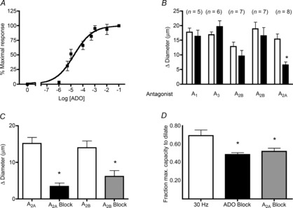
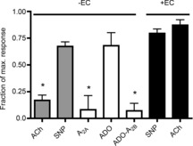
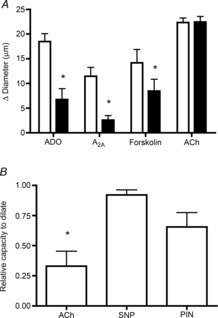
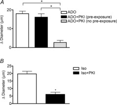
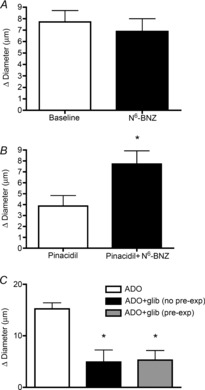
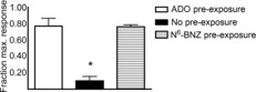
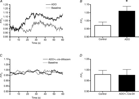
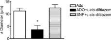
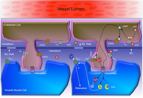
Adenosine (ADO) is an endogenous vasodilatory purine widely recognized to be a significant contributor to functional hyperaemia. Despite this, many aspects of the mechanisms by which ADO induces dilation in small resistance arterioles are not established, or appear contradictory. These include: identification of the primary receptor subtype; its location on endothelial (EC) or vascular smooth muscle cells; whether ADO acts on KATP channels in these resistance vessels; and the contribution of cAMP/protein kinase A (PKA) signalling to the response. In intravital microscopy studies of intact or EC-denuded skeletal muscle arterioles, we show that ADO acts via A2A receptors located on ECs to produce vasodilation via activation of KATP channels located on vascular smooth muscle cells. Importantly, we found that the signalling pathway involves cAMP as expected, but that a requirement for PKA activation is demonstrable only if the vessel is not pre-exposed to ADO. That is, PKA-dependent signalling varies with pre-exposure to ADO. Further, we show that PKA activation alone is not sufficient to dilate these arterioles; an additional EC calcium-dependent signalling mechanism is required for vasodilation to ADO. The ability of arterioles in situ to respond to occupancy of a specific receptor by utilizing different cell signalling pathways under different conditions to produce the same response allows the arteriole to respond to key homeostatic requirements using more than a single signalling mechanism. Clearly, this is likely to be physiologically advantageous, but the role for this signalling flexibility in the integrated arteriolar response that underlies functional hyperaemia will require further exploration.


Figures

Figure 1
Figure 1. Dilation to ADO and to muscle contraction is mediated by ADO A2A receptors
A, concentration–response curve for ADO (n = 5–7). B, A2A antagonist SCH442416 significantly attenuates the dilation to ADO, unlike the A1 antagonist DPCPX, the A3 antagonist MRS1334, and the A2B antagonists MRS1754 and PSB603, which do not. White bars, ADO; black bars, ADO+antagonist. C, dilation to the A2A agonist CGS21860 is significantly decreased in the presence of the A2A antagonist SCH442416 (black bar, n = 5), and dilation to the A2B agonist BAY 60–6583 is significantly decreased by the A2B antagonist MRS1754 (grey bar, n = 11). D, dilation induced by muscle contraction (white bar, field stimulation, 30 Hz, 15 s, n = 7) is significantly attenuated by the non-specific ADO inhibitor, xanthine amine congener (black bar, n = 5) and is equally attenuated by the A2A antagonist SCH442416 (grey bar, n = 7). Each column represents 2 min average change in diameter (A–C) or fraction of the maximum capacity to dilate (D). Bars are means ± s.e.m. *Significantly different from control response (P ≤ 0.05). ADO, adenosine.
Figure 2
Figure 2. A2A-dependent dilation requires intact endothelium
Intact blood-perfused arterioles that were denuded of their endothelium did not dilate significantly to the A2A agonist CGS21860 (white bar, n = 5), compared to the response in intact arterioles that is shown in Fig. 1. In the absence of endothelium, dilation to ADO (white bar, n = 8) was not attenuated, but was significantly attenuated by the A2B antagonist PSB603 (white bar, n = 7), which was ineffective in blocking dilation to ADO in intact arterioles (Fig. 1). Effective disruption of endothelium was confirmed by the significantly decreased dilation to ACh (grey bar, n = 8), with continuing dilation to SNP (grey bar, n = 8). Bars are means of maximal capacity to dilate, ± s.e.m. *Significantly different from mean responses to ACh and SNP in intact vessels (black bars), P ≤ 0.05. ACh, acetylcholine; ADO, adenosine; EC, endothelial cell; SNP, sodium nitroprusside.
Figure 3
Figure 3. Dilation mediated by ADO, A2A receptor activation, and by activation of adenylate cyclase, is dependent on KATP channels located on vascular smooth muscle
A, KATP channel blocker glibenclamide (black bars) significantly inhibits the dilation to ADO (n = 9), the A2A agonist CGS21860 (n = 6), and the adenylate cyclase activator forskolin (n = 7) but does not affect the dilation to ACh (n = 7), which acts via a different cell signalling pathway. *Significantly different from response in absence of glibenclamide (white bars). B, in endothelial denuded arterioles (confirmed by the significant decrease in dilation to ACh), the response to the KATP channel activator pinacidil is not significantly different from that produced by SNP (n = 9 for each condition). *Significantly different from dilation to SNP. All bars are group means ± s.e.m., P ≤ 0.05. ACh, acetylcholine; ADO, adenosine; PIN, pinacidil; SNP, sodium nitroprusside.
Figure 4
Figure 4. Pre-exposure to ADO alters the role of protein kinase A in the dilation produced by ADO
A, PKA inhibitor, PKI does not alter the dilation to ADO when the vessel was pre-exposed to ADO (black bar, n = 8) but significantly attenuates the dilation to ADO when the vessel was not pre-exposed to ADO (hatched bar, n = 6). White bar, control response to ADO (n = 8). B, after pre-exposure to isoproterenol, PKI significantly attenuates the dilation to isoproterenol (n = 6). All bars are group mean ± s.e.m. *Significantly different from control response, P ≤ 0.05. ADO, adenosine; Iso, isoproterenol; PKI, myristoylated PKI[12-22]; PKA, protein kinase A.
Figure 5
Figure 5. PKA activation alone is not sufficient to induce dilation, but can potentiate the arteriolar dilation to pinacidil. The contribution of KATP channels to the ADO dilation is not changed with ADO pre-exposure
A, PKA activator N6-BNZ (black bar) does not produce arteriolar dilation above baseline (n = 7). B, arteriolar response to pinacidil (white bar) was enhanced in the presence of the PKA activator N6-BNZ (black bar). *Significantly different from pinacidil alone (n = 7, P ≤ 0.05). C, inhibition of dilation to ADO by the KATP channel blocker glibenclamide is not different after ADO pre-exposure (grey bar) compared to no pre-exposure (black bar). n = 8. *Significantly different from ADO alone (white bar), P ≤ 0.05. All bars are group means ± s.e.m. ADO, adenosine; N6-BNZ, N6-benzoyl-cAMP; glib, glibenclamide; PKA, protein kinase A.
Figure 6
Figure 6. PKA activation is able to mimic the pre-exposure effect seen with ADO
Pre-exposure to the PKA activator N6-BNZ prevented inhibition of dilation by the PKA inhibitor PKI (hatched bar, n = 5) in the same way that pre-exposure to ADO prevented inhibition of dilation by PKI (white bar, n = 8); in contrast, with no pre-exposure to the PKA activator, PKI inhibited the dilation to ADO as expected (black bar, n = 6). *Significantly different from control response, P ≤ 0.05. All bars are means ± s.e.m. ADO, adenosine; N6-BNZ, N6-benzoyl-cAMP; PKA, protein kinase A; PKI, myristoylated PKI[12-22].
Figure 7
Figure 7. Adenosine induces an increase in EC calcium via cyclic nucleotide gated channels
A, averaged time course of EC Ca2+ (44 cells) in baseline (control) conditions (grey line) and in response to ADO (black line) in Fluo-4 loaded intact blood-perfused arterioles. EC Ca2+ changes in (A) and (C) are expressed as relative change in fluorescence intensity. B, averaged representation of the data presented in (A). C, averaged time course of EC Ca2+ (43 cells) in baseline conditions (grey line) and in response to ADO in the presence of the cyclic nucleotide gated channel blocker l-cis-diltiazem (black line). D, averaged representation of the data presented in (C). *Significantly different from control (P ≤ 0.05). Bars in (B) and (D) are means ± s.e.m. ADO, adenosine; EC, endothelial cell.
Figure 8
Figure 8. Inhibition of cyclic nucleotide gated channels attenuates ADO dilation
Dilation to ADO in the absence (white bar) or presence (black bar) of the cyclic nucleotide gated channel blocker l-cis-diltiazem. Arteriolar functionality and l-cis-diltiazem specificity is demonstrated by the ability to dilate in response to SNP in the presence of l-cis-diltiazem (striped bar). *Significantly different from ADO (P ≤ 0.05). Bars are mean of n = 6 responses, ± s.e.m. ADO, adenosine; SNP, sodium nitroprusside.
Figure 9
Figure 9. Schematic illustration of proposed model for cell signalling mechanisms underlying the dilation to ADO in small resistance arterioles in situ in skeletal muscle
The schematic shows an EC with a process projecting through one of several openings in the IEL and thus closely apposed to the underlying VSMC. The left-hand version shows the location on the plasma membranes of the receptors and channels that we studied directly (or, for the myoendothelial gap junction, inferred from our data). Clearly, other transmembrane receptors, channels and transporters are likely to contribute to the integrated functional response. The right-hand side summarizes the cell signalling mechanisms that from our data we propose link A2A receptor activation on ECs to KATP channel activation on VSMCs. Thus, ADO binds to the A2A receptor on ECs, resulting in cAMP production. cAMP then activates both CNGCs (leading to increased EC Ca2+) and via the myoendothelial gap junction, activates PKA in the VSMC. Ca2+ (or a Ca2+ activated signalling intermediate) also then passes through a myoendothelial gap junction to the VSMC. cAMP activates PKA and, separately, Ca2+ activates the as yet unidentified intermediate: both are required to activate the KATP channel and so cause VSMC relaxation. Prolonged phosphorylation of KATP channels by PKA results in the exclusion of PKA activation from subsequent responses to ADO via A2A receptors (the pre-exposure effect): A2A receptors still activate the CNGC pathway, enabling the Ca2+-dependent component of KATP channel activation to act on the phosphorylated channel to cause VSMC relaxation. AC, adenylate cyclase, ADO, adenosine; EC, endothelial cell; IEL, internal elastic lamina; CNGC, cyclic nucleotide gated channel; PKA, protein kinase A; VSMC, vascular smooth muscle cell.

Similar articles

Cited by

References

    1. Banitt PF, Smits P, Williams SB, Ganz P, Creager MA. Activation of ATP-sensitive potassium channels contributes to reactive hyperemia in humans. Am J Physiol Heart Circ Physiol. 1996;271:H1594–H1598. - PubMed
    1. Bockman EL, Berne RM, Rubio R. Adenosine and active hyperemia in dog skeletal muscle. Am J Physiol. 1976;230:1531–1537. - PubMed
    1. Carroll MA, Doumad AB, Li J, Cheng MK, Falck JR, McGiff JC. Adenosine2A receptor vasodilation of rat preglomerular microvessels is mediated by EETs that activate the cAMP/PKA pathway. Am J Physiol Renal Physiol. 2006;291:F155–F161. - PubMed
    1. Cazorla O, Lucas A, Poirier F, Lacampagne A, Lezoualc'h F. The cAMP binding protein Epac regulates cardiac myofilament function. Proc Natl Acad Sci U S A. 2009;106:14144–14149. - PMC - PubMed
    1. Cheng KT, Leung YK, Shen B, Kwok YC, Wong CO, Kwan HY, Man YB, Ma X, Huang Y, Yao X. CNGA2 channels mediate adenosine-induced Ca2+ influx in vascular endothelial cells. Arterioscler Thromb Vasc Biol. 2008;28:913–918. - PubMed

Publication types

LinkOut - more resources