Review
. 2014 Jun 15;592(12):2453-71.
doi: 10.1113/jphysiol.2014.271346. Epub 2014 Mar 31.

Polycystins and partners: proposed role in mechanosensitivity

Affiliations
Review

Polycystins and partners: proposed role in mechanosensitivity

Kevin Retailleau et al. J Physiol. .

Abstract

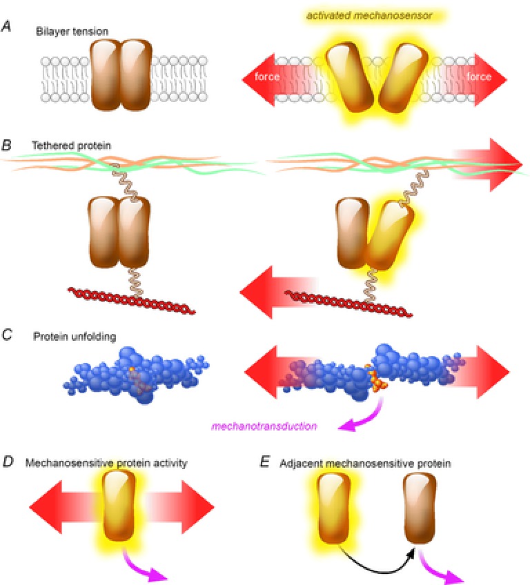
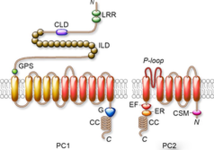
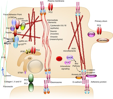
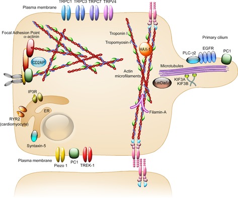
Mutations of the two polycystins, PC1 and PC2, lead to polycystic kidney disease. Polycystins are able to form complexes with numerous families of proteins that have been suggested to participate in mechanical sensing. The proposed role of polycystins and their partners in the kidney primary cilium is to sense urine flow. A role for polycystins in mechanosensing has also been shown in other cell types such as vascular smooth muscle cells and cardiac myocytes. At the plasma membrane, polycystins interact with diverse ion channels of the TRP family and with stretch-activated channels (Piezos, TREKs). The actin cytoskeleton and its interacting proteins, such as filamin A, have been shown to be essential for these interactions. Numerous proteins involved in cell-cell and cell-extracellular matrix junctions interact with PC1 and/or PC2. These multimeric protein complexes are important for cell structure integrity, the transmission of force, as well as for mechanosensing and mechanotransduction. A group of polycystin partners are also involved in subcellular trafficking mechanisms. Finally, PC1 and especially PC2 interact with elements of the endoplasmic reticulum and are essential components of calcium homeostasis. In conclusion, we propose that both PC1 and PC2 act as conductors to tune the overall cellular mechanosensitivity.


Figures

Figure 1
Figure 1. Models of proteins mechanosensitivity
Applied force is indicated by a red arrow, mechanotransduction by a magenta arrow; activated mechanosensor is highlighted in yellow. A, ‘Bilayer tension model’ with the force sensed through the lipid bilayer proposed, for example, in the bacteria mechanosensitive channel MscL (Yoshimura & Sokabe, 2010). B, ‘Tethered protein models’, with accessory proteins linking to the ECM or cytoskeleton, as described for MEC proteins in the touch sensation in Caenorhabditis elegans (Brierley, 2010). C, ‘Protein unfolding model’ with the unravelling of cryptic sites under mechanical stress, demonstrated for the extracellular matrix protein fibronectin (Gao et al. 2002). D, ‘Mechanosensitive protein activity’ with endogenous activity (enzyme, ligand, ion channel) directly activated by the perceived force, E, ‘Adjacent mechanosensitive model’ in which a primary mechanosensor indirectly activates another partner for mechanotransduction.
Figure 2
Figure 2. Predicted structures of PC1 (PKD1) (left panel) and of PC2 (TRPP2) (right panel)
The plasma membrane is depicted in grey. Abbreviations: C, C-terminus; CC, coiled-coil domain; CLD, C-lectin-like domain; CSM, ciliary sorting motif; EF, calcium-binding EF-hand domain; ER, ER retention domain; G, G protein binding site; GPS, G-protein-coupled receptor proteolytic site; ILD, Ig-like domains (PKD repeats); LRR, leucine-rich repeats; N, N-terminus.
Figure 3
Figure 3. Localisation of PC1 partners with a putative role in mechanosensing
PC1 interacts with proteins localised mostly at focal adhesion points, adherens junctions, desmosomes and in the primary cilium. Localisation is indicated in black and bold, the partners are indicated in black, and the cell is schematised in light orange.
Figure 4
Figure 4. Localisation of PC2 partners with a putative role in mechanosensing
PC2 interacts more specifically with ion channels at the plasma membrane, with proteins of the actin microfilament network and in the primary cilium. Localisation is indicated in black and bold, the partners are indicated in black, and the cell is schematised in light orange.

Similar articles

Cited by

References

    1. AbouAlaiwi WA, Takahashi M, Mell BR, Jones TJ, Ratnam S, Kolb RJ, Nauli SM. Ciliary polycystin–2 is a mechanosensitive calcium channel involved in nitric oxide signaling cascades. Circ Res. 2009;104:860–869. - PMC - PubMed
    1. Alenghat FJ, Nauli SM, Kolb R, Zhou J, Ingber DE. Global cytoskeletal control of mechanotransduction in kidney epithelial cells. Exp Cell Res. 2004;301:23–30. - PubMed
    1. Anyatonwu GI, Estrada M, Tian X, Somlo S, Ehrlich BE. Regulation of ryanodine receptor-dependent calcium signaling by polycystin–2. Proc Natl Acad Sci U S A. 2007;104:6454–6459. - PMC - PubMed
    1. Arnadottir J, Chalfie M. Eukaryotic mechanosensitive channels. Annu Rev Biophys. 2010;39:111–137. - PubMed
    1. Arnould T, Kim E, Tsiokas L, Jochimsen F, Grüning W, Chang JD, Walz G. The polycystic kidney disease 1 gene product mediates protein kinase C α–dependent and c-Jun N-terminal kinase-dependent activation of the transcription factor AP 1. J Biol Chem. 1998;273:6013–6018. - PubMed

Publication types

Substances

LinkOut - more resources