Review
. 2014 Nov;62(11):1790-815.
doi: 10.1002/glia.22658. Epub 2014 Mar 31.

Pathophysiology of glia in perinatal white matter injury

Affiliations
Review

Pathophysiology of glia in perinatal white matter injury

Stephen A Back et al. Glia. 2014 Nov.

Abstract

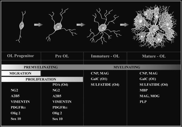
Injury to the preterm brain has a particular predilection for cerebral white matter. White matter injury (WMI) is the most common cause of brain injury in preterm infants and a major cause of chronic neurological morbidity including cerebral palsy. Factors that predispose to WMI include cerebral oxygenation disturbances and maternal-fetal infection. During the acute phase of WMI, pronounced oxidative damage occurs that targets late oligodendrocyte progenitors (pre-OLs). The developmental predilection for WMI to occur during prematurity appears to be related to both the timing of appearance and regional distribution of susceptible pre-OLs that are vulnerable to a variety of chemical mediators including reactive oxygen species, glutamate, cytokines, and adenosine. During the chronic phase of WMI, the white matter displays abberant regeneration and repair responses. Early OL progenitors respond to WMI with a rapid robust proliferative response that results in a several fold regeneration of pre-OLs that fail to terminally differentiate along their normal developmental time course. Pre-OL maturation arrest appears to be related in part to inhibitory factors that derive from reactive astrocytes in chronic lesions. Recent high field magnetic resonance imaging (MRI) data support that three distinct forms of chronic WMI exist, each of which displays unique MRI and histopathological features. These findings suggest the possibility that therapies directed at myelin regeneration and repair could be initiated early after WMI and monitored over time. These new mechanisms of acute and chronic WMI provide access to a variety of new strategies to prevent or promote repair of WMI in premature infants.

Keywords: hyperoxia; hypoxia; hypoxia-ischemia; myelin; oligodendrocyte.


Figures

Figure 1
Figure 1
Maturation of the oligodendrocyte (OL) lineage. Four principal stages of OL lineage progression are depicted together with their corresponding morphological features and capacity for myelination, migration and proliferation. Each stage is uniquely defined by a combination of marker genes or antibodies. A2B5, O4, O1 refer to mouse monoclonal antibodies. Olig 2 and Sox10 are genes that are highly enriched in premyelinating OLs. Olig 2 is also expressed at later stage in the OL lineage. Abbreviations: CNP (CNPase), 2’:3’-cyclic nucleotide-3’-phosphodiesterase; ; GalC, galactocerebroside; MAG, myelin associated glycoprotein; MBP, myelin basic protein; MOG, myelin oligodendrocyte glycoprotein; NG2, chondroitin sulfate proteoglycan 4; PDGFRα, platelet-derived growth factor-alpha; PLP, proteolipid protein.
Figure 2
Figure 2
Three forms of high field MRI-defined perinatal WMI with corresponding histopathological features that were generated in the 0.65 gestation fetal sheep brain at 1 or 2 weeks after global cerebral ischemia (adapted from: Riddle et al., 2011). Upper Panel. Diffuse WMI in chronic lesions (A) Representative appearance and distribution of diffuse hypointense (D-hypo) lesions seen on a T2w image at 1 week after injury. (B) Diffuse WMI had pronounced astrogliosis defined by immunohistochemical staining of reactive astrocytes with glial fibrillary acidic protein (GFAP; green) and a lesser population of Iba1-labeled microglia/macrophages (red) with a reactive morphology (inset). Nuclei in the inset are visualized with Hoechst 33342 (blue). (C) Quantification of GFAP-labeled astrocytes and Iba1-labeled microglia within MRI-defined WM signal abnormalities at 1 and 2 weeks after global ischemia. The D-hypo lesions had significantly elevated GFAP, consistent with a diffuse astrogliotic response to injury. * p<0.05, n.s., not significant. Bar in B, 100 μm. Middle Panel. Focal Necrotic WMI. (A) Representative appearance from the largest focal hyperintense (F-hyper) lesion seen on a T2w image at 1 week after injury. These lesions typically localized to subcortical white matter. Note the substantial difference in the F-hyper lesion relative to the diffuse gliotic lesions, which appears much more hypointense (D-hypo). (B) A typical macroscopic necrotic lesion defined by diffuse dense staining for reactive microglia and macrophages with Iba1 (red and inset) and a paucity of GFAP-labeled astrocytes. Nuclei in the inset are visualized with Hoechst 33342 (blue). (C ) F-hyper lesions displayed a progressive decrease in GFAP staining and markedly increased Iba1 labeling for microglia by 2 weeks after global ischemia. * p<0.05. Bar in B, 100 μm. Lower Panel. Microscopic necrotic WMI. (A) Representative appearance of a focal hypointense (F-hypo) lesion seen on a T2w image at 2 weeks after injury. Note the substantial difference in the F-hypo lesion relative to a diffuse gliotic lesion at 2 weeks, which appears more hyperintense (D-hyper). (B) A typical microscopic necrotic lesion defined by a discrete focus of immunohistochemical staining for reactive microglia and macrophages with Iba1 (red and inset) and a paucity of staining for astrocytes with glial fibrillary acidic protein (GFAP; green). Nuclei in the inset are visualized with Hoechst 33342 (blue). (C) F-hypo lesions had markedly increased Iba1 labeling and no significant difference in GFAP labeling vs. control. * p<0.05. Bar in B, 100 μm.
Figure 3
Figure 3
Numerous late oligodendrocyte progenitors (preOLs) accumulate in chronic myelin-deficient perinatal white matter lesions. Lesions were generated in response to unilateral hypoxia-ischemia in the postnatal day 3 rat with the contralateral hemisphere serving as control (see Segovia et al., 2008). (A) Normal early myelination (O1-antibody; green) in control subcortical white matter (corpus callosum/external capsule) at P10 is seen with low levels of GFAP-labeled astrocytes (red) mostly concentrated over the white matter. (B) Absence of myelin in the contralateral post-ischemic lesion coincided with a diffuse glial scar that stained for GFAP-labeled astrocytes. (C) Early myelination in control white matter at P10 with sheaths (yellow) double-labeled for O4 and O1 antibodies. (D) Absence of myelin in the contralateral lesion coincided with clusters of preOLs (O4+O1-) in maturation arrest (red; arrowheads). Such dense clusters of preOLs are not normally seen in control white matter and are consistent with the pronounced proliferative state that is triggered in response to injury. Peak preOL density can expand roughly 4-fold relative to control. Oligodendroctes (yellow; arrows; O4+O1+) are rarely seen in the lesions. Abbreviations: CTX, cerebral cortex; CPu, caudate putamen.

References

    1. (CDC) CfDCaP. Economic costs associated with mental retardation, cerebral palsy, hearing loss and vision impairment--United States, 2003. MMWR Morb Mortal Wkly Rep. 2004;53:57–59. - PubMed
    1. Aarnoudse-Moens CS, Weisglas-Kuperus N, van Goudoever JB, Oosterlaan J. Meta-analysis of neurobehavioral outcomes in very preterm and/or very low birth weight children. Pediatrics. 2009;124:717–28. - PubMed
    1. Agresti C, D'Urso D, Levi G. Reversible inhibitory effects of interferon-γ and tumor necrosis factor-α on oligodendroglial lineage cell proliferation and differentiation in vitro. Eur J Neurosci. 1996;8:1106–1116. - PubMed
    1. Aguirre A, Dupree JL, Mangin JM, Gallo V. A functional role for EGFR signaling in myelination and remyelination. Nat Neurosci. 2007;10:990–1002. - PubMed
    1. Alix JJ, Fern R. Glutamate receptor-mediated ischemic injury of premyelinated central axons. Ann Neurol. 2009;66:682–93. - PubMed

Publication types

Substances