. 2014 May 1;192(9):4453-4464.
doi: 10.4049/jimmunol.1400146. Epub 2014 Mar 31.

Immunological priming requires regulatory T cells and IL-10-producing macrophages to accelerate resolution from severe lung inflammation

Affiliations

Immunological priming requires regulatory T cells and IL-10-producing macrophages to accelerate resolution from severe lung inflammation

Neil R Aggarwal et al. J Immunol. .

Erratum in

Abstract

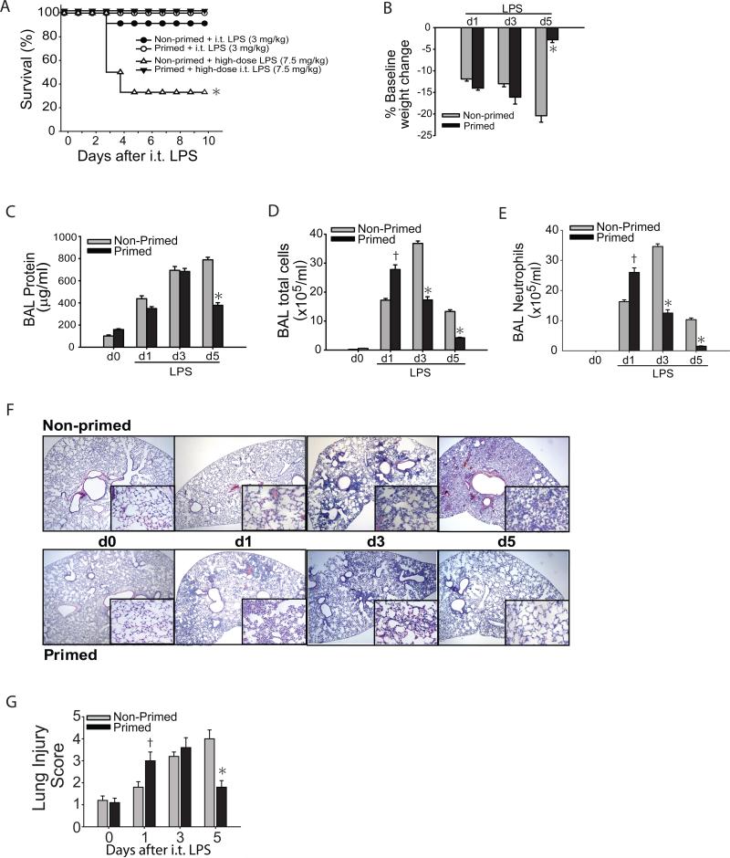
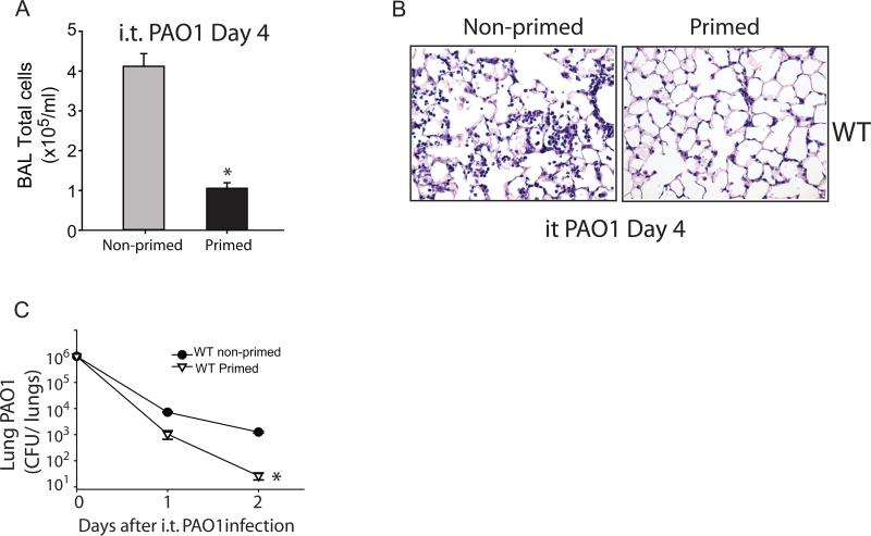
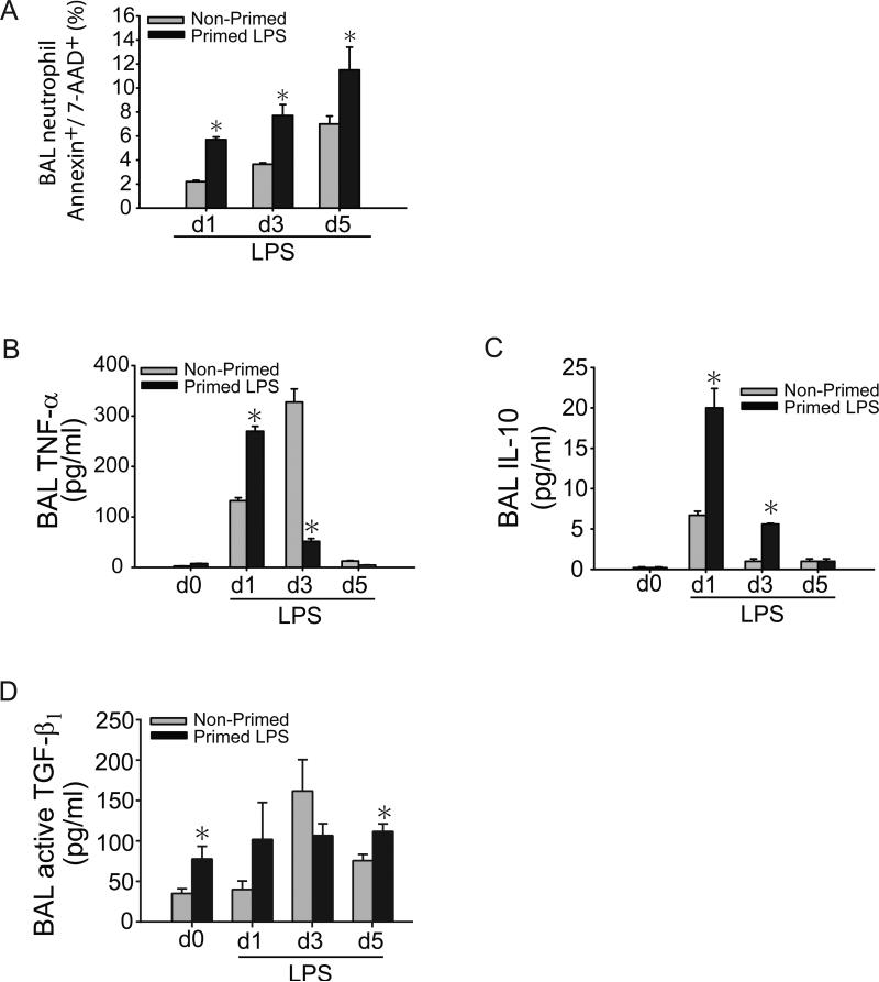
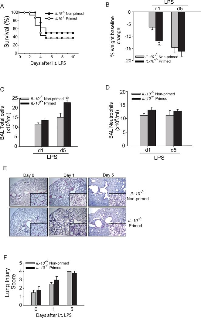
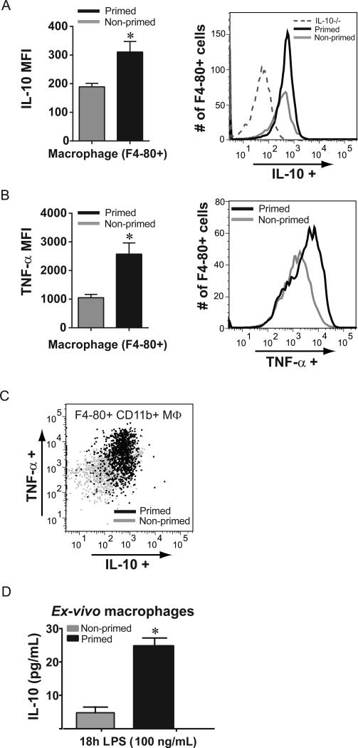
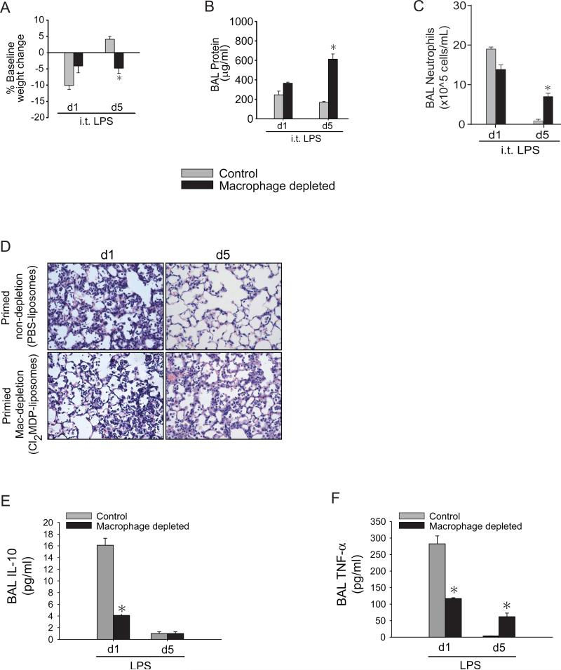
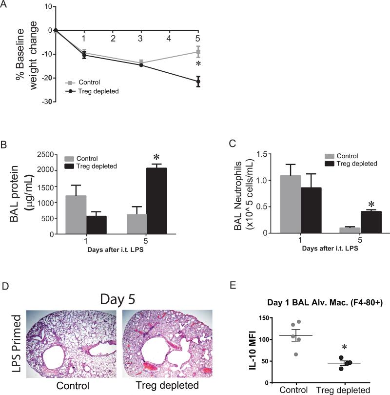
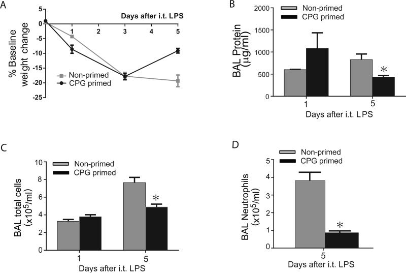
Overwhelming lung inflammation frequently occurs following exposure to both direct infectious and noninfectious agents and is a leading cause of mortality worldwide. In that context, immunomodulatory strategies may be used to limit severity of impending organ damage. We sought to determine whether priming the lung by activating the immune system, or immunological priming, could accelerate resolution of severe lung inflammation. We assessed the importance of alveolar macrophages, regulatory T cells, and their potential interaction during immunological priming. We demonstrate that oropharyngeal delivery of low-dose LPS can immunologically prime the lung to augment alveolar macrophage production of IL-10 and enhance resolution of lung inflammation induced by a lethal dose of LPS or by Pseudomonas bacterial pneumonia. IL-10-deficient mice did not achieve priming and were unable to accelerate lung injury resolution. Depletion of lung macrophages or regulatory T cells during the priming response completely abrogated the positive effect of immunological priming on resolution of lung inflammation and significantly reduced alveolar macrophage IL-10 production. Finally, we demonstrated that oropharyngeal delivery of synthetic CpG-oligonucleotides elicited minimal lung inflammation compared with low-dose LPS but nonetheless primed the lung to accelerate resolution of lung injury following subsequent lethal LPS exposure. Immunological priming is a viable immunomodulatory strategy used to enhance resolution in an experimental acute lung injury model with the potential for therapeutic benefit against a wide array of injurious exposures.


Figures

Fig. 1
Fig. 1. Immunological priming accelerates lung injury resolution
Following a 7-day priming period, primed and non-primed WT mice were assessed for survival after either 3 mg/kg or 7.5 mg/kg i.t. LPS (A). After either dose of i.t. LPS, survival over 10 days was determined in primed and non-primed WT mice (n=8-10 per time point). Primed and non-primed WT mice were assessed for body weight relative to baseline (B), bronchoalveolar (BAL) protein (C), BAL total cell counts (D) and BAL neutrophils (E) at intervals after i.t. LPS injury. (F) Histological sections were stained with H & E in primed and non-primed WT mice. Original magnifications x20; x100 (inserts). (G) Histopathological mean lung injury scores from x20 sections (n=4-6 animals per group per time point). Values expressed as mean ± SEM; * or † paired t-test against other group at same time point, p<0.05, * log-rank test for survival curve.
Fig. 2
Fig. 2. Immunological priming accelerates resolution from bacterial pneumonia
Following an 7-day priming period, BAL total cells (A) and lung histology by H&E staining (B) were assessed in primed and non-primed WT mice at day 4 after i.t. PAO1 (1×10⁶ CFU). Bacterial clearance was determined by measurement of whole lung PAO1 colony-forming units (CFU) in WT primed and non-primed mice on days 1 and 2 after i.t. PAO1 (C). Values expressed as mean ± SEM; *paired t-test against other group at same time point, p<0.05. (n=4-6 animals per group per time point)
Fig. 3
Fig. 3. Lung priming modulates the alveolar inflammatory milieu
(A) Apoptosis (Annexin V+/7-AAD+) of BAL neutrophils from primed and non-primed WT mice on days 1, 3, or 5 after i.t. LPS was assessed by flow cytometry. BAL neutrophils were gated by characteristic granulocyte forward and side scatter, sub-gated for Gr-1+ to identify neutrophils, and then for AnnexinV/7-AAD, percentages for which are quantified in (A). BAL TNF-α (B), IL-10 (C), and active TGF-β1 (D) cytokine secretion were assessed at designated time points after i.t. LPS in primed and non-primed WT mice. Values expressed as mean ± SEM; *paired t-test against other group at same time point, p<0.05. (n=4-6 animals per group per time point)
Fig. 4
Fig. 4. IL-10−/− mice do not benefit from immunological priming
(A) Survival was determined in primed and non-primed IL-10−/− mice. Primed and non-primed IL-10−/− mice were assessed for body weight relative to baseline (B), BAL total cell counts (C) or BAL neutrophils (D) at days 1 or 5 after i.t. LPS injury. (E) Histological sections were stained with H & E in primed and non-primed WT mice. Original magnifications x20; x100 (inserts). (F) Histopathological mean lung injury scores from x20 sections. Values expressed as mean ± SEM; *paired t-test against other group at same time point, p<0.05, (n=4-6 animals per group per time point). log-rank test for survival curve, n=8-10 in primed and non-primed groups.
Fig. 5
Fig. 5. Macrophages are a significant IL-10 source after lung priming
Alveolar cells from primed and non-primed WT mice were isolated at day +1 after i.t. LPS (3 mg/kg) challenge, and then restimulated, and stained for macrophage, neutrophil, and lymphocyte flow markers as well as intracellular cytokine production. IC IL-10 production (A) and IC TNF-α production (B) were quantified by mean fluorescence intensity (MFI) in F4-80+ alveolar macrophages collected from primed and non-primed WT mice; a representative flow cytometry histogram is shown for each, and for IL-10 includes alveolar macrophages from primed IL-10−/− mice (dashed line). (C) Among F4-80+ CD11b+ alveolar cells from primed (black) and non-primed (gray) mice, a dot plot demonstrating individual cell IC production demonstrates a predominant increase in dual cytokine production from primed alveolar cells. (D) Alveolar macrophages were isolated 7 days after o.p. LPS or o.p. water (control), and stimulated with LPS (100 ng/mL); IL-10 secretion was quantified by ELISA after 18 hours of stimulation. Values expressed as mean ± SEM; *paired t-test against other group at same time point, p<0.05. (n=4-5 animals or wells per group per time point)
Fig. 6
Fig. 6. IL-10 producing alveolar macrophages are critical for immunological priming
Primed mice treated with PBS-liposomes (control) and primed mice treated with CL2-MDP liposomes (macrophage depleted) were assessed for body weight relative to baseline (A), BAL total protein (B), BAL neutrophils (C) or histological damage (D) by H&E staining (x100 magnification) at days 1 or 5 after i.t. LPS injury. BAL IL-10 (E) and TNF-α from primed control and primed macrophage-depleted were measured at days 1 or 5 after i.t. LPS. Values expressed as mean ± SEM; *one-way ANOVA (A-C) or paired t-test (E-F) against other groups at same time point, p<0.05, (n=4-6 animals per group per time point)
Fig. 7
Fig. 7. Tregs are necessary for priming and alveolar macrophage IL-10 production
Primed Foxp3gfp mice (control) and primed Foxp3DTR (Treg depleted) were assessed for body weight relative to baseline (A), BAL total protein (B), BAL neutrophils (C), or histological damage (D) by H&E staining (x2x magnification) at days 1 or 5 after i.t. LPS injury. All mice received i.p. DT injections (15 μg/kg) on days −2, −1, and +2 when harvested at day 5. (E) At day 1 after i.t. LPS, we determined intracellular production of IL-10 among F4-80+ BAL macrophages by flow cytometry. Values expressed as mean ± SEM, as well as individual values in E; *paired t-test against other primed group at same time point, p<0.05, (n=4-6 animals per group per time point)
Fig. 8
Fig. 8. Immunological priming is achieved with minimal inflammation
WT mice were treated with o.p. PBS (Non-primed) or o.p. CpG (primed). 5 days later, mice were challenged with i.t. LPS (3 mg/kg), and were assessed for body weight relative to baseline (A), BAL total protein (B), BAL total cells (C), or BAL neutrophils (D) at days 1 or 5 after i.t. LPS injury. Values expressed as mean ± SEM; *paired t-test against other primed group at same time point, p<0.05, (n=4-6 animals per group per time point)

References

    1. Romero CD, Varma TK, Hobbs JB, Reyes A, Driver B, Sherwood ER. The Toll-like receptor 4 agonist monophosphoryl lipid a augments innate host resistance to systemic bacterial infection. Infect Immun. 2011;79:3576–3587. - PMC - PubMed
    1. Meldrum DR, Cleveland JC, Jr., Moore EE, Partrick DA, Banerjee A, Harken AH. Adaptive and maladaptive mechanisms of cellular priming. Ann Surg. 1997;226:587–598. - PMC - PubMed
    1. Ariel A, Maridonneau-Parini I, Rovere-Querini P, Levine JS, Muhl H. Macrophages in inflammation and its resolution. Front Immunol. 2012;3:324. - PMC - PubMed
    1. Bem RA, Farnand AW, Wong V, Koski A, Rosenfeld ME, van Rooijen N, Frevert CW, Martin TR, Matute-Bello G. Depletion of resident alveolar macrophages does not prevent Fas-mediated lung injury in mice. Am J Physiol Lung Cell Mol Physiol. 2008;295:L314–325. - PMC - PubMed
    1. Doherty DE, Downey GP, Worthen GS, Haslett C, Henson PM. Monocyte retention and migration in pulmonary inflammation. Requirement for neutrophils. Lab Invest. 1988;59:200–213. - PubMed

Publication types