

Reproducibility of peripapillary retinal nerve fiber layer thickness measured by spectral domain optical coherence tomography in pseudophakic eyes
- PMID: 24688256
- PMCID: PMC3958629
- DOI: 10.3341/kjo.2014.28.2.138
Reproducibility of peripapillary retinal nerve fiber layer thickness measured by spectral domain optical coherence tomography in pseudophakic eyes
Abstract
Purpose: To assess the reproducibility of circumpapillary retinal nerve fiber layer (cpRNFL) thickness measurement (measurement agreement) and its color-coded classification (classification agreement) by Cirrus spectral domain optical coherence tomography (OCT) in pseudophakic eyes.
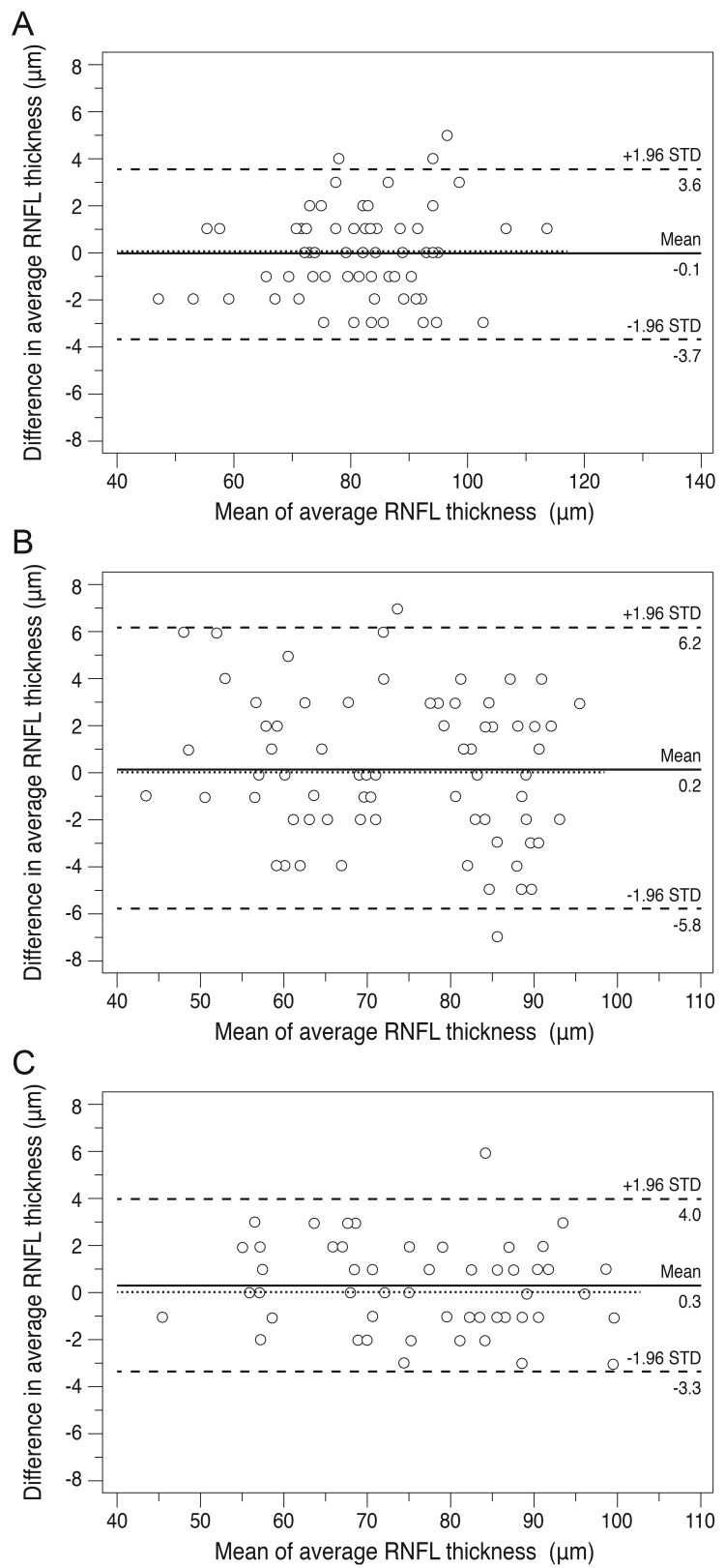
Methods: Two-hundred five participants having glaucoma or glaucoma suspected eyes underwent two repeated Cirrus OCT scans to measure cpRNFL thickness (optic disc cube 200 × 200). After classifying participants into three different groups according to their lens status (clear media, cataract, and pseudophakic), values of intra-class coefficient (ICC), coefficient of variance, and test-retest variability were compared between groups for average retinal nerve fiber layer (RNFL) thicknesses and that corresponding to four quadrant maps. Linear weighted kappa coefficients were calculated as indicators of agreement of color code classification in each group.
Results: ICC values were all excellent (generally defined as 0.75 to 1.00) for the average and quadrant RNFL thicknesses in all three groups. ICC values of the clear media group tended to be higher than those in the cataract and pseudophakic groups for all quadrants and average thickness. Especially in the superior and nasal quadrants, the ICC value of the cataract group was significantly lower than that of the clear media and pseudophakic groups. For average RNFL thickness, classification agreement (kappa) in three groups did not show a statistically significant difference. For quadrant maps, classification agreement (kappa) in the clear media group was higher than those in the other two groups.
Conclusions: Agreement of cpRNFL measurement and its color code classification between two repeated Cirrus OCT scans in pseudophakic eyes was as good as that in eyes with clear crystalline lens. More studies are required to ascertain the effect of lens status on the reproducibility of Cirrus OCT according to different stages of glaucoma patients.
Keywords: Optical coherence tomography; Pseudophakia; Reproducibility of results.
Conflict of interest statement
No potential conflict of interest relevant to this article was reported.
Figures
References
-
- Carpineto P, Nubile M, Agnifili L, et al. Reproducibility and repeatability of Cirrus HD-OCT peripapillary retinal nerve fibre layer thickness measurements in young normal subjects. Ophthalmologica. 2012;227:139–145. - PubMed
-
- Cremasco F, Massa G, Goncalves Vidotti V, et al. Intrasession, intersession, and interexaminer variabilities of retinal nerve fiber layer measurements with spectral-domain OCT. Eur J Ophthalmol. 2011;21:264–270. - PubMed
-
- Pareja-Esteban J, Teus-Guezala MA, Drake-Casanova P, Dapena-Sevilla I. Retinal nerve fiber layer changes after cataract surgery measured by OCT: a pilot study. Arch Soc Esp Oftalmol. 2009;84:305–309. - PubMed
-
- El-Ashry M, Appaswamy S, Deokule S, Pagliarini S. The effect of phacoemulsification cataract surgery on the measurement of retinal nerve fiber layer thickness using optical coherence tomography. Curr Eye Res. 2006;31:409–413. - PubMed
-
- Mwanza JC, Bhorade AM, Sekhon N, et al. Effect of cataract and its removal on signal strength and peripapillary retinal nerve fiber layer optical coherence tomography measurements. J Glaucoma. 2011;20:37–43. - PubMed
Publication types
MeSH terms
LinkOut - more resources
Full Text Sources
Other Literature Sources
Medical
