

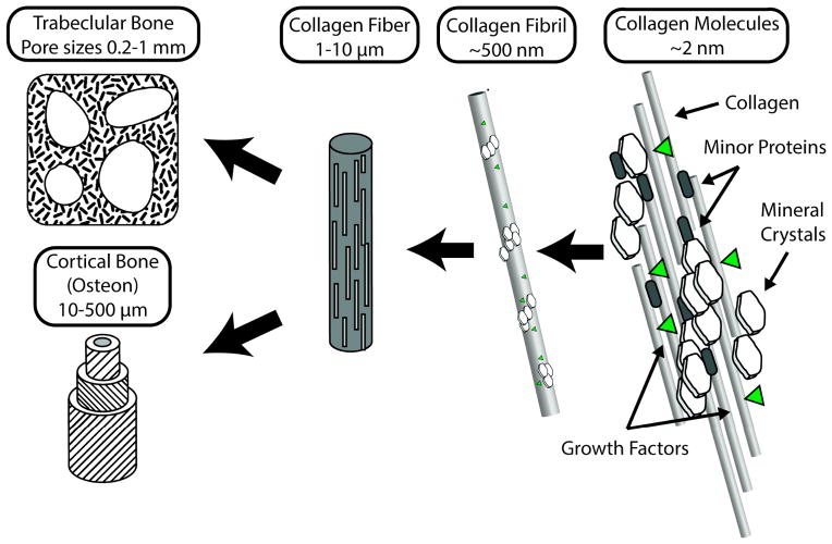
Mimicking the nanostructure of bone matrix to regenerate bone
- PMID: 24688283
- PMCID: PMC3968917
- DOI: 10.1016/j.mattod.2013.11.001
Mimicking the nanostructure of bone matrix to regenerate bone
Abstract
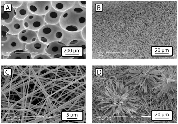
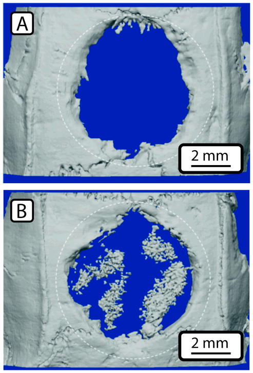
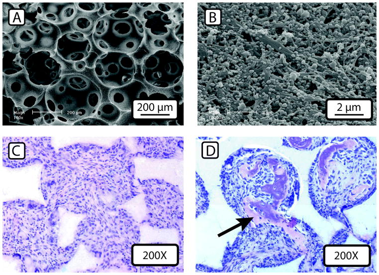
Key features of bone tissue structure and composition are capable of directing cellular behavior towards the generation of new bone tissue. Bone tissue, as well as materials derived from bone, have a long and successful history of use as bone grafting materials. Recent developments in design and processing of synthetic scaffolding systems has allowed the replication of the bone's desirable biological activity in easy to fabricate polymeric materials with nano-scale features exposed on the surface. The biological response to these new tissue-engineering scaffold materials oftentimes exceeds that seen on scaffolds produced using biological materials.
Keywords: Biomimetic; Bone tissue engineering; Nanofiber; Scaffold; Thermally induced phase separation.
Figures
References
Grants and funding
LinkOut - more resources
Full Text Sources
Other Literature Sources