

Role of ultrasound in the understanding and management of vasculitis
- PMID: 24688604
- PMCID: PMC3956137
- DOI: 10.1177/1759720X13512256
Role of ultrasound in the understanding and management of vasculitis
Abstract
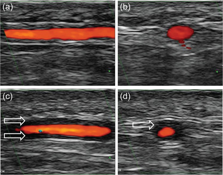
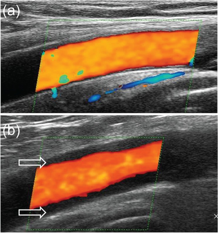
Vasculitis is characterized by a circumferential vessel-wall thickening ('halo'), which can be visualized by modern imaging techniques. In particular, the resolution of ultrasound has increased to 0.1 mm. Ultrasound detects abnormalities that are pathognomonic even in arteries with a diameter below 1 mm. It is particularly helpful in the diagnosis of large-vessel vasculitides, such as classic temporal arteritis, large-vessel giant-cell arteritis (GCA), Takayasu arteritis and idiopathic aortitis. Echocardiography is important for determining cardiac involvement in Takayasu arteritis and also for examining the coronary arteries of children with suspected Kawasaki disease, which is a medium-vessel vasculitis. In small vessel vasculitides ultrasound has only a role for determining the distribution or organ involvement. Fast-track clinics for the diagnosis of GCA help to initiate treatment before complications such as blindness occur; patients receive appointments within 24 h in these clinics. Clinical examination and ultrasound of temporal and axillary arteries are performed by an experienced rheumatologist. In most cases this is able to determine if GCA is present. Temporal artery biopsy can be still carried out in ambivalent cases. The wall swelling of temporal arteries disappears after 2-3 weeks of glucocorticoid treatment. After 3 days of treatment, diagnosis becomes more difficult with ultrasound in some cases. In larger arteries, such as the axillary arteries, wall thickening disappears within months. It tends to be darker (more hypoechoic) in acute disease because of oedema.
Keywords: Kawasaki disease; Takayasu arteritis; aortitis; giant-cell arteritis; temporal arteritis; ultrasound; vasculitis.
Conflict of interest statement
Figures
References
-
- Aschwanden M., Daikeler T., Kesten F., Baldi T., Benz D., Tyndall A., et al. (2013) Temporal artery compression sign – a novel ultrasound finding for the diagnosis of giant cell arteritis. Ultraschall Med 34: 47–50 - PubMed
-
- Baer A., Rubin L., Shapiro C., Sood S., Rajan S., Shapir Y., et al. (2006) Prevalence of coronary artery lesions on the initial echocardiogram in Kawasaki syndrome. Arch Pediatr Adolesc Med 160: 686–690 - PubMed
-
- Ball E., Walsh S., Tang T., Gohil R., Clarke J. (2010) Role of ultrasonography in the diagnosis of temporal arteritis. Br J Surg 97: 1765–1771 - PubMed
Publication types
LinkOut - more resources
Full Text Sources
Other Literature Sources
