Review
. 2012 Nov 9:2:e201209015.
doi: 10.5936/csbj.201209015. eCollection 2012.

Cellulose degradation by oxidative enzymes

Affiliations
Review

Cellulose degradation by oxidative enzymes

Maria Dimarogona et al. Comput Struct Biotechnol J. .

Abstract

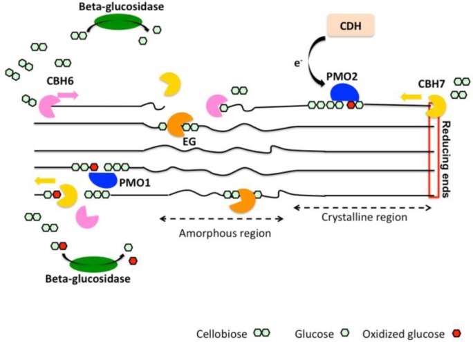
Enzymatic degradation of plant biomass has attracted intensive research interest for the production of economically viable biofuels. Here we present an overview of the recent findings on biocatalysts implicated in the oxidative cleavage of cellulose, including polysaccharide monooxygenases (PMOs or LPMOs which stands for lytic PMOs), cellobiose dehydrogenases (CDHs) and members of carbohydrate-binding module family 33 (CBM33). PMOs, a novel class of enzymes previously termed GH61s, boost the efficiency of common cellulases resulting in increased hydrolysis yields while lowering the protein loading needed. They act on the crystalline part of cellulose by generating oxidized and non-oxidized chain ends. An external electron donor is required for boosting the activity of PMOs. We discuss recent findings concerning their mechanism of action and identify issues and questions to be addressed in the future.

Keywords: CBM33; GH61; bioethanol; biofuels; cellulose; polysaccharide monooxygenases.


Figures

Figure 1
Figure 1
A. The figure shows the structure of Cel61B (molB, PDB code 2VTC) in cartoon representation. Conserved residues on the surface of the molecule are shown in ball and stick representation. B. The nickel ion (purple sphere) coordinated by His1, His 89, Tyr 176 and two water molecules (red spheres) in Cel61B structure. C. The structure of CBP21 (molC, PDB code 2BEM) in cartoon representation. Highlighted in ball and stick are the highly conserved residues His114 and His28, and a bound sodium ion (blue sphere). D. The sodium ion (blue sphere) coordinated by His28 and His 114 in molC of CBP21 structure. All figures were prepared with Molsoft [27].
Figure 2
Figure 2
Oxidized reaction products released from GH61s applied on cellulosic substrates.
Figure 3
Figure 3
A. Superposition of the crystal structures of PMO-2 (PDB code 4EIR, molA) in orange and PMO-3 (PDB code 4EIS, molA) in cyan. Loop L2 is highlighted by a dashed square and the additional glycosylation site on PMO-3 is shown in ball and stick representation. B. Copper coordination site of PMO-3. The methylation site on the N-terminal histidine is shown in a dashed square. C. Multiple sequence alignment of PMO enzymes categorized in three types according to sequence, structural and biochemical characteristics (based on [37, 41]: Type 1: NCU08760 (NcPMO-1), PcGH61D, TtGH61E, type 2: NCU01050 (NcPMO-2), NCU02916(GH61-3) and type 3: NCU07898 (NcPMO-3), TaGH61A. Blue asterisks mark copper-coordinating residues. The secondary structure elements shown in blue and disulphide bonds (numbers in green) were assigned based on the crystal structure of NcPMO-3 (PDB code 4EIS). Residues in the orange frame form loop L2. Identical and similar residues are printed in white on a red background and in red on a white background, respectively. Secondary structure elements α-helices, 310-helices, β-strands and strict α and β-turns are denoted as α, η, β, TTT and TT, respectively. Multiple sequence alignment of homologous enzymes was performed with Clustal Omega [42] at EBI server and visualized with ESPript 2.2 [43]. Figures were prepared with Molsoft.
Figure 4
Figure 4
Schematic representation of a CDH comprising a C-terminal flavin domain with its FAD highighted in green (PDB code 1NAA, [51]) and an N-terminal heme domain (PDB code 1D7B, [52]. The protein structures were visualized with Molsoft [27]. Oligosaccharide oxidation takes place at the flavin domain followed by electron transfer to the ferric heme group.
Figure 5
Figure 5
A simplified scheme of the current view on the enzymatic degradation of cellulose, involving cellobiohydrolases (CBH), endoglucanases (EG), type1 and type 2 PMOs (PMO1 and PMO2, respectively). Cellobiose dehydrogenase (CDH) is a potential electron donor for PMOs. EGs and PMOs cleave internally cellulose chains releasing chain ends that are targeted by CBHs. CBHs generate cellobiose or oxidized cellobiose that are subsequently hydrolyzed by β-glucosidase.

References

    1. Panwar NL, Kaushik S. C, Kothari Surendra (2011) Role of renewable energy sources in environmental protection: A review. Renewable and Sustainable Energy Reviews 15: 1513–1524
    1. Energy USDo (2010) Biomass basics: The facts about bioenergy.
    1. Sims, RE, Mabee, W, Saddler, JN, Taylor, M (2010) An overview of second generation biofuel technologies. Bioresour Technol 101: 1570–1580 - PubMed
    1. Sun, Y, Cheng, J (2002) Hydrolysis of lignocellulosic materials for ethanol production: a review. Bioresour Technol 83: 1–11 - PubMed
    1. Himmel, ME, Ding, SY, Johnson, DK, Adney, WS, Nimlos, MR,et al. . (2007) Biomass recalcitrance: engineering plants and enzymes for biofuels production. Science 315: 804–807 - PubMed

LinkOut - more resources