Review
. 2013 Sep 3:6:e201303010.
doi: 10.5936/csbj.201303010. eCollection 2013.

The essential biology of the endoplasmic reticulum stress response for structural and computational biologists

Affiliations
Review

The essential biology of the endoplasmic reticulum stress response for structural and computational biologists

Sadao Wakabayashi et al. Comput Struct Biotechnol J. .

Abstract

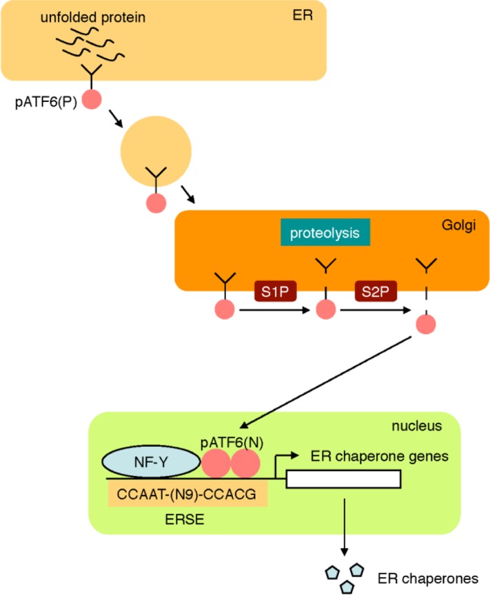
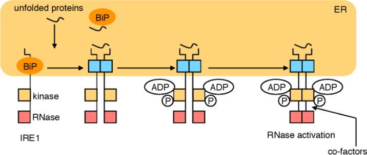
The endoplasmic reticulum (ER) stress response is a cytoprotective mechanism that maintains homeostasis of the ER by upregulating the capacity of the ER in accordance with cellular demands. If the ER stress response cannot function correctly, because of reasons such as aging, genetic mutation or environmental stress, unfolded proteins accumulate in the ER and cause ER stress-induced apoptosis, resulting in the onset of folding diseases, including Alzheimer's disease and diabetes mellitus. Although the mechanism of the ER stress response has been analyzed extensively by biochemists, cell biologists and molecular biologists, many aspects remain to be elucidated. For example, it is unclear how sensor molecules detect ER stress, or how cells choose the two opposite cell fates (survival or apoptosis) during the ER stress response. To resolve these critical issues, structural and computational approaches will be indispensable, although the mechanism of the ER stress response is complicated and difficult to understand holistically at a glance. Here, we provide a concise introduction to the mammalian ER stress response for structural and computational biologists.

Keywords: ATF6; ER stress; ERAD; IRE1; PERK; XBP1; unfolded protein response.


Figures

Figure 1
Figure 1
The ER stress response. The ER stress response is a cytoprotective mechanism that regulates the expression of ER chaperones as well as ERAD factors at the transcriptional level in accordance with cellular demands. When unfolded proteins accumulate in the ER, sensor molecules activate specific transcription factors, leading to transcriptional activation of genes encoding ER chaperones and ERAD factors. The ER stress response is thought to wane with aging, resulting in ER stress-induced apoptosis of cells and finally various diseases, which are collectively called “folding diseases”.
Figure 2
Figure 2
The ATF6 pathway. The sensor molecule pATF6(P) located on the ER membrane is transported to the Golgi apparatus by transport vesicles in response to ER stress. In the Golgi apparatus, pATF6(P) is sequentially cleaved by two proteases, S1P and S2P, resulting in release of the cytoplasmic portion pATF6(N) from the ER membrane. pATF6(N) translocates into the nucleus and activates transcription of ER chaperone genes through binding to the cis-acting enhancer ERSE.
Figure 3
Figure 3
The IRE1 pathway. In normal growth conditions, the sensor molecule IRE1 is an inactive monomer, whereas IRE1 forms an active oligomer in response to ER stress. Activated IRE1 converts unspliced XBP1 mRNA to mature mRNA by the cytoplasmic mRNA splicing. From mature XBP1 mRNA, an active transcription factor pXBP1(S) is translated and activates the transcription of ERAD genes through binding to the enhancer UPRE.
Figure 4
Figure 4
The PERK pathway. When PERK detects unfolded proteins in the ER, PERK phosphorylates eIF2α, resulting in translational attenuation and translational induction of ATF4. ATF4 activates the transcription of target genes encoding translation factors, anti-oxidation factors and a transcription factor CHOP. Other kinases such as PKR, GCN2 and HRI also phosphorylate eIF2α, and phosphorylated eIF2α is dephosphorylated by CReP, PP1C-GADD34 and p58IPK.
Figure 5
Figure 5
Working hypothesis of the activation mechanism of IRE1. In normal growth conditions, BiP binds to an IRE1 monomer and prevents IRE1 from forming an oligomer. Upon ER stress, BiP binds to unfolded proteins, and IRE1 forms an oligomer, to which unfolded proteins directly bind and cause a conformational change in IRE1. Then, ADP binds to IRE1 and IRE1 is transphosphorylated, resulting in activation of its RNase. Hypothetical cytosolic co-factors may bind to the cytoplasmic ligand-binding pocket and may modulate IRE1 activity.
Figure 6
Figure 6
Two working models of the activation mechanism of ATF6. According to the first model, BiP binds to pATF6(P) and masks the Golgi localization signal in normal growth conditions. Upon ER stress, unfolded proteins sequester BiP from pATF6(P), resulting in translocation of pATF6(P) to the Golgi apparatus. In the second model, pATF6(P) forms an oligomer through disulfide bonds and is anchored in the ER membrane, while disulfide bonds are cleaved by reduction upon ER stress and a pATF6(P) monomer translocates to the Golgi apparatus. Both models are not mutually exclusive.
Figure 7
Figure 7
Three functions of pXBP1(U). pXBP1(U) translated from XBP1(U) mRNA binds to pXBP1(S) and enhances its degradation. The CTR region of pXBP1(U) interacts with the ribosome tunnel and slows translation, while the HR2 region anchors XBP1(U) mRNA to the ER membrane, in order to enhance splicing of XBP1(U) mRNA by IRE1.
Figure 8
Figure 8
Major pathways of ER stress-induced apoptosis. ER stress induces apoptosis through various pathways, including transcriptional induction of CHOP by the PERK and ATF6 pathways, the IRE1-TRAF2 pathway and the caspase-12 pathway.

References

    1. Gething M-J (1997) Guidebook to Molecular Chaperones and Protein-Folding Catalysts. Oxford: Oxford University Press
    1. Hebert DN, Molinari M (2012) Flagging and docking: dual roles for N-glycans in protein quality control and cellular proteostasis. Trends Biochem Sci 37: 404–410 - PMC - PubMed
    1. Tabas I, Ron D (2011) Integrating the mechanisms of apoptosis induced by endoplasmic reticulum stress. Nat Cell Biol 13: 184–190 - PMC - PubMed
    1. Imaizumi K, Miyoshi K, Katayama T, Yoneda T, Taniguchi M, et al. (2001) The unfolded protein response and Alzheimer's disease. Biochim Biophys Acta 1536: 85–96 - PubMed
    1. Wang HQ, Takahashi R (2007) Expanding insights on the involvement of endoplasmic reticulum stress in Parkinson's disease. Antioxid Redox Signal 9: 553–561 - PubMed

LinkOut - more resources