. 2013 Dec 15:6:e201303022.
doi: 10.5936/csbj.201303022. eCollection 2013.

Of mice and men: Dissecting the interaction between Listeria monocytogenes Internalin A and E-cadherin

Affiliations

Of mice and men: Dissecting the interaction between Listeria monocytogenes Internalin A and E-cadherin

Samuel Genheden et al. Comput Struct Biotechnol J. .

Abstract

We report a study of the interaction between internalin A (inlA) and human or murine E-cadherin (Ecad). inlA is used by Listeria monocytogenes to internalize itself into host cell, but the bacterium is unable to invade murine cells, which has been attributed to the difference in sequence between hEcad and mEcad. Using molecular dynamics simulations, MM/GBSA free energy calculations, hydrogen bond analysis, water characterization and umbrella sampling, we provide a complete atomistic picture of the binding between inlA and Ecad. We dissect key residues in the protein-protein interface and analyze the energetics using MM/GBSA. From this analysis it is clear that the binding of inlA-mEcad is weaker than inlA-hEcad, on par with the experimentally observed inability of inlA to bind to mEcad. However, extended MD simulations of 200 ns in length show no destabilization of the inlA-mEcad complex and the estimation of the potential of mean force (PMF) using umbrella sampling corroborates this conclusion. The binding strength computed from the PMFs show no significant difference between the two protein complexes. Hence, our study suggests that the inability of L. monocytogenes to invade murine cells cannot be explained by processes at the nanosecond to sub-microsecond time scale probed by the simulations performed here.

Keywords: E-cadherin; Internalin A; energy decomposition; molecular dynamics; umbrella sampling.


Figures
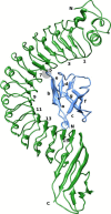
Figure 1
Figure 1
Complex between inlA in green and hEcad in blue. The numbering of every second inlA β-sheets(LRRs) is shown, as well as the numbering of the β-sheets in hEcad. N– and C–termini of the protein chains are marked with an N and C, respectively. The hEcad loop containing Pro16 and the tip of LRR 6 is encircled in grey.
Figure 2
Figure 2
Illustration of mutation in inlA, a) and b), and differences between hEcad and mEcad, c) and d). a) Illustrates the S192N mutation, which leads to an interaction between Asn192 and Phe17. b) Illustrates the Y369S mutation, which leads to an interaction with Asn27. c) Illustrates the important Pro/Glu16 difference between hEcad and mEcad. d) Illustrates the Glu/Gln64 difference.
Figure 3
Figure 3
Illustration of the direction of displacement in the umbrella calculations. inlA is shown in green at one edge of the simulation box. The position of hEcad as observed in the crystal structure is then shown in blue, and at displacements of 24 Å and 48 Å in purple and pink, respectively. The simulation box is sketched for reference. The hEcad loop containing Pro16 and the tip of LRR 6 is encircled in grey.
Figure 4
Figure 4
Free energy contributions of residues on inlA and Ecad in kJ/mol. a) inlA residues in the inlA–hEcad complex, b) inlA residues in the inlA–mEcad complex, c) Ecad residues in the inlA–hEcad complex, d) Ecad residues in the inlA–mEcad complex. Residues were selected based on a number of criteria as outlined in the text. Free energy contributions are determined by energy decomposition (ED), alanine scanning mutagenesis (ASM), and scaled ASM (sASM).
Figure 5
Figure 5
Per-residue free energy difference between wild-type inlA–Ecad complex and various mutants. a) Difference relative to inlA–hEcad complex for residues on inlA, b) Difference relative to inlA–hEcad complex for residues on Ecad, c) Difference relative to inlA–mEcad complex for residues on inlA, d) Difference relative to inlA–mEcad complex for residues on Ecad
Figure 6
Figure 6
Conserved water sites in the interface. Showing the location of the sites listed in Table VI. The green protein is inlA and the blue protein is Ecad. Sites are shown as red and orange spheres, the red were found for the inlA–hEcad complex and the orange for the inlA–mEcad complex. Residues with 3 Å of the sites are shown as well.
Figure 7
Figure 7
Average PMF for inlA–hEcad (blue) and inlA–mEcad (red).
Figure 8
Figure 8
Illustration of the two main clusters of hot spots. inlA is shown in green and hEcad is shown in blue, hot spots are colored by atom.

Similar articles

Cited by

References

    1. Bierne H, Cossart P (2007) Listeria monocytogenes surface proteins: from genome predictions to function. Microbiol mol biol rev 71: 377–397 - PMC - PubMed
    1. Vazquez-Boland JA, Kuhn M, Berche P, Chakraborty T, Dominguez-Bernal G, et al. (2001) Listeria Pathogenesis and Molecular Virulence Determinants. Clinic Microbiol Rev 14: 584–640 - PMC - PubMed
    1. Bonazzi M, Lecuit M, Cossart P (2009) Listeria monocytogenes internalin and E-cadherin: from bench to bedside. Cold Spring Harb Perspect Biol 1: a003087. - PMC - PubMed
    1. Hamon M, Bierne H, Cossart P (2006) Listeria monocytogenes: a multifaceted model. Nat rev Microbiol 4: 423–434 - PubMed
    1. Bierne H, Sabet C, Personnic N, Cossart P (2007) Internalins: a complex family of leucine-rich repeat-containing proteins in Listeria monocytogenes. Microbes Infect 9: 1156–1166 - PubMed

LinkOut - more resources