. 2013 Sep 10:7:e201304006.
doi: 10.5936/csbj.201304006. eCollection 2013.

Biochemical and biophysical characterization of recombinant yeast proteasome maturation factor ump1

Affiliations

Biochemical and biophysical characterization of recombinant yeast proteasome maturation factor ump1

Bebiana Sá-Moura et al. Comput Struct Biotechnol J. .

Abstract

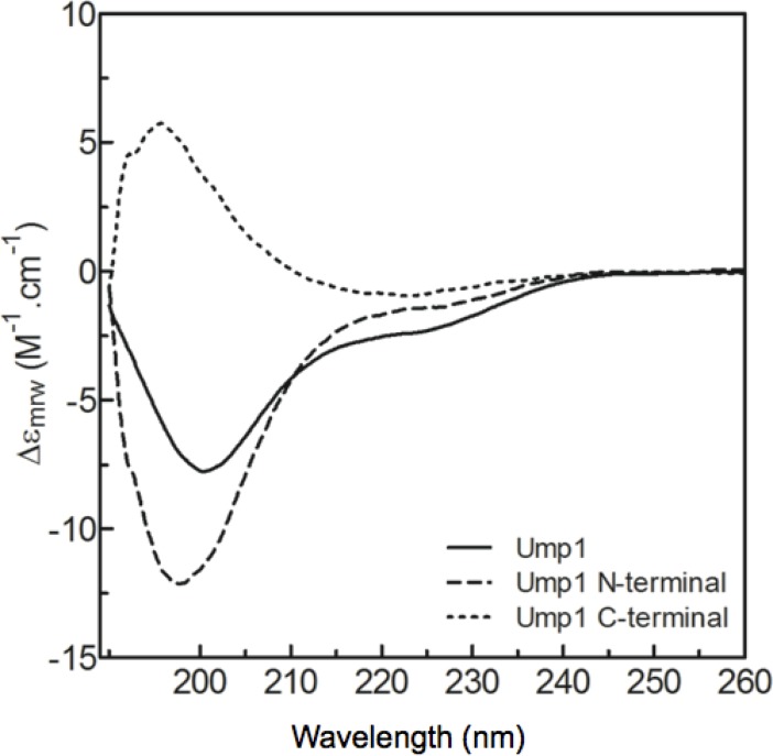
Protein degradation is essential for maintaining cellular homeostasis. The proteasome is the central enzyme responsible for non-lysosomal protein degradation in eukaryotic cells. Although proteasome assembly is not yet completely understood, a number of cofactors required for proper assembly and maturation have been identified. Ump is a short-lived maturation factor required for the efficient biogenesis of the 20S proteasome. Upon the association of the two precursor complexes, Ump is encased and is rapidly degraded after the proteolytic sites in the interior of the nascent proteasome are activated. In order to further understand the mechanisms behind proteasomal maturation, we expressed and purified yeast Ump in E. coli for biophysical and structural analysis. We show that recombinant Ump is purified as a mixture of different oligomeric species and that oligomerization is mediated by intermolecular disulfide bond formation involving the only cysteine residue present in the protein. Furthermore, a combination of bioinformatic, biochemical and structural analysis revealed that Ump shows characteristics of an intrinsically disordered protein, which might become structured only upon interaction with the proteasome subunits.

Keywords: Circular dichroism; dynamic light scattering; intrinsically disordered; protein structure.


Figures

Figure 1
Figure 1
Recombinant Ump1 is purified as a mixture of molecular species with different charges and oligomeric states. A) Ion-exchange chromatographic profile of the metal-affinity purified Ump1 fraction shows that this protein is further separated into two peaks corresponding to species with different isoelectric points (peak 1 and peak 2). Conductivity is represented by a dotted line. B) Electrophoretic analysis of Ump1 fractions corresponding to peak 1 (monomer) and peak 2 (dimer) of the ion-exchange chromatography. The wild-type Ump1 monomer is frequently contaminated with dimers under non-reducing conditions (first lane). The Ump1-C115S mutant elutes from the ion-exchange column as a single peak (data not shown) and migrates as the wild-type Ump1 monomer. Proteins were loaded in sample buffer without (-) or with (+) 10 mM DTT prior to electrophoresis in a 15% SDS-PAGE (here stained with Coomassie Blue). MW, Molecular weight marker; values in kDa.
Figure 2
Figure 2
The N-terminal half of Ump1 is predicted to be highly disordered. Amino acid sequence alignment of selected Ump1 proteins (see Table S1 for protein% similarities) was performed with ClustalW2, and rendered with Aline [35]. Disorder was predicted with RONN [30] for the selected amino acid sequences and a consensus line for disorder prediction (http://www.bioinformatics.nl/∼berndb/ronn.html) is printed below the alignment: the black line highlights residues where disorder is predicted for all the displayed sequences and the blue line represents regions where disorder is predicated for least 80% of the represented Ump1 orthologs. The position of the non-conserved Cys115 is indicated by a red star, the conserved motif HPLE is indicated by red triangles, and the Cys37 residue conserved in mammalian orthologs is boxed. The blue-boxed arrow above Arg84 points to one of the trypsin-cleavage sites identified by N-terminal sequencing after limited proteolysis experiments (Figure S1). SCHCE, Ump1 from Saccharomyces cerevisiae (UniProt accession code P38293); SCHPO, Ump1 from Schizosaccharomyces pombe (O74416); MOUSE, Ump1 from Mus musculus (Q9CQT5); PONAB, Ump1 from Pongo abelii (Q5R9L9); HUMAN, Ump1 from Homo sapiens (Q9Y244); BOVIN, Ump1 from Bos taurus (Q3SZV5); DICDI, Ump1 ortholog from Dictyostelium discoideum (Q55G18) and DRMEG, Ump1 from Drosophila melanogaster (Q9VIJ5).
Figure 3
Figure 3
Determination of Ump1 apparent molecular mass and Stokes radii (Rs). A) Size-exclusion chromatography of wild-type Ump1 dimer and Ump1-C115S monomer. Superdex 75 calibration was performed with the following molecular weight protein standards: 1 - aprotinin (6.5 kDa), 2 – ribonuclease A (13.7 kDa), 3 - chymotrypsinogen (25.0 kDa), and 4 - ovalbumin (43.0 kDa). Ump1 wild-type dimer and C115S monomer display atypical mobility, eluting with apparently higher molecular masses of 65 and 40 kDa, respectively (calculated with the equation Kav = -2.0693•log(MW) + 4.9698, R2 = 0.99607, obtained after column calibration). Using these data, the apparent Rs calculated for wild-type Ump1 dimer and C115S monomer are 34 and 27 Å, respectively (as calculated from the equation Rs = 0.3467(1000/Ve)-5.7834, R2 = 0.99061; Ve = elution volume). B) Logarithmic plot of Rs versus molecular mass (MW) of the corresponding proteins. The straight lines represent the average theoretical Rs for the proteins used as standards, assuming different conformational states (native), a natively unfolded pre-molten globule-like conformation (nu-PMG) or a non-native urea-denatured conformation (un) according to the equations given in ref [26]. The error bars represent the standard deviation for each plot as calculated from ref. [26]. Ump1 monomer (C115S) and Ump1 dimer correspond to the orange and red circles, respectively and fall within the range of values expected for natively unfolded molten globule conformation. For comparison purposes, experimentally determined values for Rs [36] are shown for pre-molten globule conformations of proteins with molecular masses of 43 kDa (MMP-1 Interstitial collagenase, orange triangle), 28 kDa (Tryptophan synthase, blue circle) and 15 kDa (Tumor suppressor p16, blue rhombus).
Figure 4
Figure 4
Analysis of Ump1 secondary structure and conformational stability by circular dichroism. A) Far-UV CD spectra of wild-type (monomer and dimer) and mutant Ump1. Monomeric wild-type Ump1 was prepared in the presence or absence of freshly prepared 1mM DTT. The strong negative peak at 201 nm is characteristic of abundant random coil structures. B) Denaturation curves were computed for wild-type (monomer and dimer, both in the presence and absence of DTT) and Ump1-C115S from the CD variation at 205 nm ellipticity (mdeg). Higher (i.e. less negative) signals indicate lower structural content and unfolding. Non-cooperative unfolding and resistance to full denaturation are characteristic of proteins harbouring unstructured segments [38].
Figure 5
Figure 5
The N-terminal region of Ump1 is highly disordered. Far-UV CD spectra of Ump1-C115S and its isolated N-terminal fragment. The difference spectrum for the C-terminal peptide was obtained by subtracting the N-terminal Ump1 spectrum from that of full-length Ump1-C115S. Upon deconvolution the secondary structure of Ump1 N-terminal is 1% α-helix, 25% β-strand, 22% turns and 50% coil. The C-terminal peptide secondary structure corresponds to 18% α-helix, 37% β-strand, 19% turns and 27% coil.

References

    1. Marques AJ, Palanimurugan R, Matias AC, Ramos PC, Dohmen RJ (2009) Catalytic mechanism and assembly of the proteasome. Chem Rev 109: 1509–1536 - PubMed
    1. Finley D (2009) Recognition and processing of ubiquitin-protein conjugates by the proteasome. Annu Rev Biochem 78: 477–513 - PMC - PubMed
    1. Lasker K, Forster F, Bohn S, Walzthoeni T, Villa E, et al (2012) Molecular architecture of the 26S proteasome holocomplex determined by an integrative approach. Proc Natl Acad Sci U S A 109: 1380–1387 - PMC - PubMed
    1. Beck F, Unverdorben P, Bohn S, Schweitzer A, Pfeifer G, et al (2012) Near-atomic resolution structural model of the yeast 26S proteasome. Proc Natl Acad Sci U S A 109: 14870–14875 - PMC - PubMed
    1. Lander GC, Estrin E, Matyskiela ME, Bashore C, Nogales E, et al (2012) Complete subunit architecture of the proteasome regulatory particle. Nature 482: 186–191 - PMC - PubMed

LinkOut - more resources