

Review
doi: 10.5936/csbj.201308003.
eCollection 2013.
A Sieve-Raft Hypothesis for the regulation of endothelial fenestrations
Affiliations
- PMID: 24688743
- PMCID: PMC3962122
- DOI: 10.5936/csbj.201308003
Item in Clipboard
Review
A Sieve-Raft Hypothesis for the regulation of endothelial fenestrations
Comput Struct Biotechnol J.
.
No abstract available
Figures
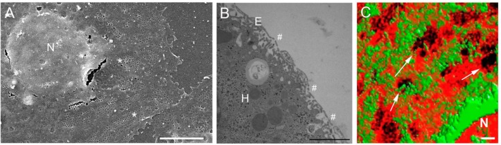
Microscopy of LSEC fenestrations and the LSEC membrane. Figure 1A is a scanning electron micrograph of an isolated LSEC in culture. The micrograph clearly displays fenestrations, examples are denoted by an asterix (*), arranged in groups (sieve plates) or individually. The fenestrations are located in the thin cytoplasmic extensions of the cell, distal to the nucleus (N) Scale bar = 5 µm. Figure 1B is a transmission electron micrograph of perfusion fixed liver, the unique architecture of the sinusoid can be seen. The very thin endothelium (E) is perforated with fenestrations (#), allowing passage of substrates into the hepatocytes (H) for metabolism, storage and detoxification Scale bar = 2 µm. Figure 1C is a micrograph prepared by 3D structured illumination microscopy. The LSECs have been stained with Bodipy FL C5 ganglioside GM1, a marker for rafts (green) and Cell-Mask Orange, a cell membrane marker (orange). There is an inverse distribution between liver sieve plates and membrane rafts. Some sieve plates are identified by an arrow (→) and fenestrations can be resolved within the sieve plates. Scale bar = 1 µm.
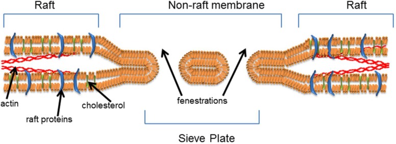
The Sieve-Raft hypothesis: the composition and arrangement of lipids in the cell membrane is paramount in determining fenestration formation and cell function. We propose that fenestrations form in non-raft microdomains of the lipid bilayer and that rafts and actin engender membrane stability, while limiting fenestration formation.
Similar articles
-
The Relationship between fenestrations, sieve plates and rafts in liver sinusoidal endothelial cells.PLoS One. 2012;7(9):e46134. doi: 10.1371/journal.pone.0046134. Epub 2012 Sep 24. PLoS One. 2012. PMID: 23029409 Free PMC article.
-
Cystathionine-Gamma-Lyase-Derived Hydrogen Sulfide-Regulated Substance P Modulates Liver Sieve Fenestrations in Caecal Ligation and Puncture-Induced Sepsis.Int J Mol Sci. 2019 Jun 29;20(13):3191. doi: 10.3390/ijms20133191. Int J Mol Sci. 2019. PMID: 31261857 Free PMC article.
-
The liver sieve and atherosclerosis.Pathology. 2012 Apr;44(3):181-6. doi: 10.1097/PAT.0b013e328351bcc8. Pathology. 2012. PMID: 22406487 Review.
-
Hyperlipidemia and surfactants: the liver sieve is a link.Atherosclerosis. 2006 Dec;189(2):273-81. doi: 10.1016/j.atherosclerosis.2005.12.025. Epub 2006 Feb 2. Atherosclerosis. 2006. PMID: 16458315
-
The wHole Story About Fenestrations in LSEC.Front Physiol. 2021 Sep 13;12:735573. doi: 10.3389/fphys.2021.735573. eCollection 2021. Front Physiol. 2021. PMID: 34588998 Free PMC article. Review.
Cited by
-
Manipulating fenestrations in young and old liver sinusoidal endothelial cells.Am J Physiol Gastrointest Liver Physiol. 2019 Jan 1;316(1):G144-G154. doi: 10.1152/ajpgi.00179.2018. Epub 2018 Oct 4. Am J Physiol Gastrointest Liver Physiol. 2019. PMID: 30285464 Free PMC article.
-
Discontinuities in the endothelium of epiphyseal cartilage canals and relevance to joint disease in foals.J Anat. 2016 Jan;228(1):162-75. doi: 10.1111/joa.12391. Epub 2015 Oct 15. J Anat. 2016. PMID: 26471892 Free PMC article.
-
Fenestrated Endothelial Cells across Organs: Insights into Kidney Function and Disease.Int J Mol Sci. 2024 Aug 22;25(16):9107. doi: 10.3390/ijms25169107. Int J Mol Sci. 2024. PMID: 39201792 Free PMC article. Review.
-
Biophysical nanocharacterization of liver sinusoidal endothelial cells through atomic force microscopy.Biophys Rev. 2020 Jun;12(3):625-636. doi: 10.1007/s12551-020-00699-0. Epub 2020 May 18. Biophys Rev. 2020. PMID: 32424787 Free PMC article. Review.
-
Actin-spectrin scaffold supports open fenestrae in liver sinusoidal endothelial cells.Traffic. 2019 Dec;20(12):932-942. doi: 10.1111/tra.12700. Epub 2019 Oct 23. Traffic. 2019. PMID: 31569283 Free PMC article.
References
-
- Hilmer, SN, Cogger VC, Fraser R, McLean AJ, Sullivan D and Le Couteur DG, Age-related changes in the hepatic sinusoidal endothelium impede lipoprotein transfer in the rat. Hepatology, 2005. 42(6): p. 1349–1354 - PubMed
-
- Pradel, G and Frevert U, Malaria sporozoites actively enter and pass through rat Kupffer cells prior to hepatocyte invasion. Hepatology, 2001. 33(5): p. 1154–1165 - PubMed
-
- Fraser, RLe Couteur DGWarren ACogger VC, Bertolino P and Smith M. The liver sieve and gene therapy. Blood,, 101(8): p. 3338; author reply 3338-3339 - PubMed
-
- Mueller, T, Rumpel E, Hradetzky S, Bollig F, Wegner H, Blumenthal A, Greinacher A, Endlich K, Endlich N. 2011. Non-muscle myosin IIA is required for the development of the zebrafish glomerulus. Kidney International, 2003. 80(10): p. 1055–1063 - PubMed
Publication types
LinkOut - more resources
Full Text Sources
Other Literature Sources